
Page 41 - 2020_09-Haematologica-web
P. 41
Molecular heterogeneity of PK deficiency
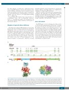
the entire lifespan of erythrocytes.13 Pathological muta- tions causing PK deficiency can be localized in any of the protein domains, with major clusters in specific regions, such as the interface between the A and C domains, the A/A′ intersubunit interface, the hydrophobic core of the A domain, and the fructose 1,6 bisphosphate-binding site6,10,14,15 (Figure 1C).
Several human PK mutants have been produced as recombinant forms and biochemically character- ized10,11,13,16,17 showing that amino acid substitutions can affect thermostability, catalytic efficiency, and response to the allosteric effector.
Diagnosis of pyruvate kinase deficiency
The diagnostic workup for PK deficiency is based on the patient’s personal and family medical history and clinical examination, and on several laboratory investigations, including the spectrophotometric assay of red blood cell PK activity.18 Molecular analysis of the PKLR gene is nec- essary to confirm the diagnosis, and overcomes the limita- tions of the enzymatic test, which may give false positive results in the case of heterozygous carriers, or false nega- tive results in the case of recent transfusion, or an increased number of reticulocytes. Therefore, recently
A
B
C
published guidelines and recommendations conclude that enzyme analyses and DNA studies are complementary techniques for diagnosing PK deficiency.19
With the advent of next-generation sequencing (NGS) techniques, the PKLR gene is usually included in panels designed for diagnosing hereditary hemolytic anemias,20-24 allowing detection of an increasing number of cases, thus reducing misdiagnosis, and highlighting the extreme phe- notypic variability of PK deficiency25-27 (Table 1).
Gene and variants
The PKLR gene, located on chromosome 1q21, consists of 12 exons and is approximately 9.5 kb in size.31 The gene encodes for the liver (L) and erythrocyte (R) isoforms of the enzyme according to tissue-specific promoters;31,32 ten exons are shared by the two isoforms, while exons 1 and 2 are specifically transcribed to the PK-R and PK-L mRNA, respectively. The cDNA encoding PK-R is 2060 bp long and codes for 574 amino acids (Figure 1A). In the R-type promoter region, two CAC boxes and four GATA motifs are located within 270 bp from the translational initiation codon; the proximal 120 bp region has basal promoter activity and the region from -120 to -270 works as a pow- erful enhancer in erythroid cells.31
Figure 1. PKLR gene and red cell pyruvate kinase structure. (A) The PKLR gene, its chromosomal localization, extension and intron/exon organization. Numbering and mutations are usually reported in the literature using the RPK cDNA sequence of the PKLR gene, with the A of the initiation ATG being assigned number +1 (Transcript refseq ID NM_000298.5). (B) Structural domains of the human PK-R monomer, the N-terminal domain is reported in yellow, A-domain in red, B-domain in light blue and C-domain in green. The corresponding amino acids are reported below. *Represents the localization of residues directly involved in the allosteric site and catalytic center (yellow) and in the fructose 1,6 bisphosphate (FBP) activator (red). (C) Ribbon representation of the human erythrocyte pyruvate kinase monomer (left) in complex with the substrate and the allosteric activator fructose-1,6-diphosphate (red and purple) and tetramer based on the crystal structure described by Valentini et al.10 Circles indicate the A’A’ and the A/C subunit interfaces.
haematologica | 2020; 105(9)
2219
