
Page 110 - 2020_09-Haematologica-web
P. 110
J. Zhou et al.
AC
B
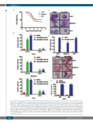
Figure 1. Effects of ASLAN003 on cell viability and differentiation in human acute myeloid leukemia cell lines. (A) Dose-response curves of ASLAN003 treatment for 48 h on cell viability of the acute myeloid leukemia (AML) cell lines THP-1, MOLM-14, and KG-1. The percentage of cell viability relative to that of dimethylsulfoxide (DMSO)-treated cells is shown. Data represent three independent replicates [mean ± standard deviation (SD)]. (B) FACS analysis of myeloid differentiation cell sur- face antigens CD11b and CD14 on ASLAN003-treated and DMSO-treated AML cell lines. The treatment time was 96 h. Data represent the mean ± SD of three repli- cates. **P<0.01; *P<0.05. (C) Representative images of Wright-Giemsa staining for morphological examination of AML cell lines treated with 100 nM ASLAN003 or DMSO for 96 h. The images were taken under an Olympus IX71 light microscope (Japan) with original magnification x 400 (objective lenses x 40). (D) Nitro blue tetrazolium (NBT) reduction assays for AML cells treated with 100 nM ASLAN003 or DMSO for 96 h. Cells positive for NBT-reducing activity, containing precipitated formazan particles, were counted. The bar graph shows the mean percentage of NBT+ cells in ten random 10 x (objective lenses) fields ± SD for each group. *P<0.001. (E) FACS analysis of CD11b+ cells after exposure of THP-1 cells to ASLAN003 or DMSO for 24 h and 48 h. The data display the mean ± SD of three different experiments. *P<0.001.
D
E
2288
haematologica | 2020; 105(9)
