
Page 280 - Haematologica Atlas of Hematologic Cytology
P. 280
ABAB
CDCD
EF
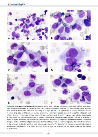
Figure 11 Amelanotic melanoma Bone marrow smear from a a a a a a a a a 30-year-old woman with fever diffuse bone pain significant hepatomegaly and and splenomegaly and and leukoerythroblastic anemia (A) Hypercellular bone marrow with an an infiltrate of of of blast cells cells often occurring as as clumps (B and C) At higher magnification these cells cells are of of of medium-large size and and have loose nuclear chromatin prominent nucleoli abundant basophilic cytoplasm and and often indistinct cell cell cell outlines (D) Few giant cells cells are multinucleated (E and and F) Polygonal and and spindle-shaped cells cells are also observed Immunocytochemistry showed that atypical cells did not express CD45 antigen or other lym- phoid or or myeloid markers nor cytokeratins they instead expressed S100 protein and the the melanoma-associated antigens melanA and the the the antigen antigen recognized by the the the antibody HMB-45 Morphologically the the the cells of amelanotic melanoma melanoma cannot be distinguished from other neoplastic cells The detection of melanoma-associated antigens allows a a a a a a a a a a a correct diagnosis to be be made It should be be noted that not not infrequently metastatic malignant melanoma is amelanotic 267
