
Page 279 - Haematologica Atlas of Hematologic Cytology
P. 279
CHAPTER 34 - Metastases of solid tumors
AB
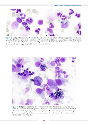
Figure 9 Malignant melanoma (A and B) Buffy coat smear from a a a a a a a a a a a patient with severe pancytopenia showing granules of of melanic pigment in the cytoplasm of of (A) a a a a a a a neutrophil and and (B) a a a a a a a monocyte monocyte Neutrophils and and monocytes can phagocytize the the the melanin deposits released by melanoma cells infiltrating the the the bone marrow and carry them into circulation thus suggesting that the bone marrow is affected Figure Figure 10 Malignant melanoma Bone marrow smear from the same case as as Figure Figure 9 demon- strates the presence of some large cells with round nuclei prominent nucleoli and abundant cytoplasm containing many very fine black granules Melanin is also present in in in in in macrophages Ten years before the patient had undergone surgery for for cutaneous melanoma thus a a a a a a a bone marrow relapse was diagnosed 266
