
Page 190 - Haematologica Atlas of Hematologic Cytology
P. 190
Chapter 20 CONGENITAL DYSERYTHROPOIETIC ANEMIAS
Congenital dyserythropoietic anemias
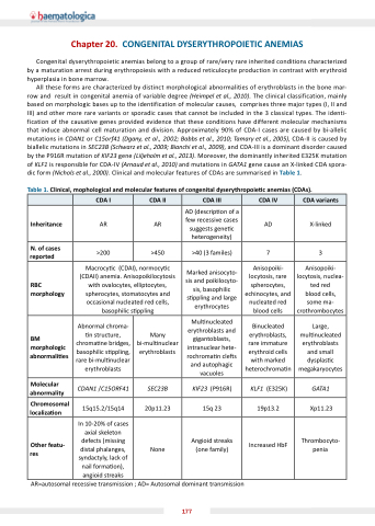
belong to a a a a a a a a group of rare/very rare rare inherited conditions characterized by a a a a a a maturation arrest during erythropoiesis with with a a a a a a reduced reticulocyte production in in contrast with with erythroid hyperplasia in bone marrow All these forms are characterized by distinct morphological abnormalities of erythroblasts in in the the bone mar- row and result in in in congenital anemia of variable degree (Heimpel et al al al 2010) The clinical classification mainly based on on morphologic bases up to the identification of molecular causes comprises three major types (I II and III) and other more rare variants or or or sporadic cases that cannot be included in in the the 3 classical types The identi- fication of the the causative genes provided evidence that these conditions have different molecular mechanisms that induce abnormal cell maturation and division Approximately 90% of CDA-I cases are caused by bi-allelic mutations in CDAN1 or or C15orf41 (Dgany et et et al al al 2002 Babbs et et et al al al 2010 Tamary et et et al al al 2005) CDA-II is caused by biallelic mutations in in SEC23B (Schwarz et et al al al 2009 2009 Bianchi et et al al al 2009) and CDA-III is is a a a a a a a a a a dominant disorder caused by the the P916R mutation mutation of KIF23 gene (Liljeholm et al 2013) Moreover the the dominantly inherited E325K mutation mutation of KLF1 is responsible for CDA-IV (Arnaud et al 2010) and mutations in in GATA1 gene cause an an X-linked CDA CDA spora- dic form (Nichols et al al 2000) Clinical and molecular features of CDAs are summarised in in Table 1 Table 1 linical mophological and molecular features of congenital dyserythropoie c c c c c anemias
( D s) CDA I CDA II CDA III CDA IV CDA variants Inheritance
AR
AR
AD (descrip on of a few recessive cases suggests gene c heterogeneity)
AD X-linked N of cases reported
>200
>450
>40 (3 familes)
7 3 RBC morphology
Macrocy c c c c c (CDAI) normocy c c c c c (CDAII) anemia Anisopoikilocytosis with ovalocytes elliptocytes spherocytes stomatocytes and occasional nucleated red cells basophilic s s ppling
Marked anisocyto- sis and poikilocyto- sis basophilic s ppling
and large erythrocytes
Anisopoiki- locytosis rare spherocytes echinocytes and nucleated red blood cells Anisopoiki- locytosis nuclea- ted red blood cells some ma- crothrombocytes
BM morphologic abnormali es Abnormal chroma- n structure chroma ne bridges basophilic s s ppling
rare bi-mul nuclear erythroblasts Many bi-mul nuclear erythroblasts Mul nucleated erythroblasts and gigantoblasts intranuclear hete- rochroma n cle s and autophagic vacuoles
Binucleated erythroblasts rare immature erythroid cells with marked heterochroma n Large mul nucleated erythroblasts and small dysplas c megakaryocytes
Molecular abnormality
CDAN1 /C15ORF41
SEC23B KIF23 (P916R)
KLF1 (E325K)
GATA1 Chromosomal localiza on 15q15 2/15q14
20p11 23 15q 23 19p13 2 Xp11 23 Other featu- res In 10-20% of cases axial skeleton defects (missing distal phalanges syndactyly lack of nail forma on) angioid streaks
None
Angioid streaks
(one family)
Increased HbF
Thrombocyto- penia AR=autosomal recessive transmission transmission AD= Autosomal dominant transmission transmission 177
