
Page 157 - Haematologica Atlas of Hematologic Cytology
P. 157
CHAPTER 17 - - Mature B-cell neoplasms
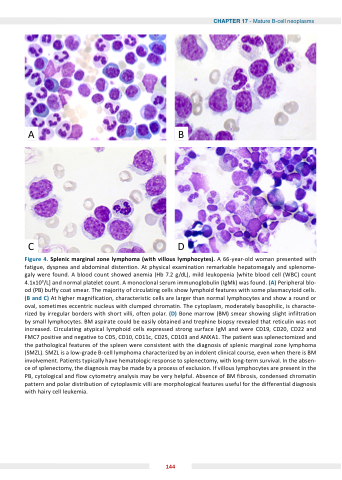
CD Figure 4 Splenic marginal zone lymphoma (with villous lymphocytes) A 66-year-old woman presented with with fatigue dyspnea and and abdominal distention At physical examination remarkable hepatomegaly and and splenome- galy were found A blood blood count count showed anemia (Hb 7 2 g/dL) mild leukopenia [white blood blood cell (WBC) count count 4 1x10⁹/L] and normal platelet count A A monoclonal serum immunoglobulin (IgMk) was found (A) Peripheral blo- od (PB) buffy coat smear The majority of circulating cells cells show lymphoid features with some plasmacytoid cells cells (B and and C) At higher magnification characteristic cells are larger than normal lymphocytes and and show a a a a a a a a a a a round or or oval sometimes eccentric nucleus with clumped chromatin The cytoplasm moderately basophilic is characte- rized by irregular borders with short villi often polar (D) Bone marrow (BM) smear showing slight infiltration by small lymphocytes BM aspirate could be easily obtained and trephine biopsy revealed that reticulin was not increased Circulating atypical lymphoid cells expressed strong surface IgM and and were CD19 CD20 CD22 and and FMC7 positive and and and negative to to CD5 CD10 CD10 CD11c CD25 CD103 and and and ANXA1 The patient was splenectomized and and and the the the pathological features of of the the the spleen were consistent with the the the diagnosis of of splenic marginal zone lymphoma (SMZL) SMZL SMZL is is a a a a a a a low-grade B-cell lymphoma characterized by an indolent clinical course even when there is is BM involvement Patients typically have hematologic response to to to splenectomy with long-term survival In the absen- ce ce of of splenectomy the the diagnosis may be made by a a a a a process of of exclusion If villous lymphocytes are present in the the PB cytological and flow cytometry analysis may be very helpful Absence of BM fibrosis condensed chromatin pattern and polar distribution of cytoplasmic villi are morphological features useful for the differential diagnosis with hairy cell leukemia 144
