
Page 156 - Haematologica Atlas of Hematologic Cytology
P. 156
ABAB
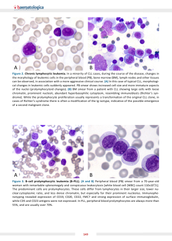
Figure 2 Chronic lymphocytic leukemia In a a a a a minority of of CLL cases during the the course of of the the disease changes in in in the the the morphology of leukemic cells in the the the peripheral blood (PB) bone marrow (BM) lymph nodes and other tissues can be observed in in association with a a a a a a a a more aggressive clinical course (A) In this case of typical CLL morphologi- cal changes in in leukemic cells suddenly appeared PB smear shows increased cell cell size and more immature aspects of the nuclei (prolymphocytoid changes) (B) BM smear from a a a a a patient with with CLL showing large cells with with loose chromatin prominent nucleoli abundant hyperbasophilic cytoplasm resembling immunoblasts (Richter’s syn- drome) While the the prolymphocyte proliferation usually represents a a a a a a transformation of the the original CLL clone in in cases of of of of Richter’s syndrome there is is often a a a a modification of of of of the the the Ig isotype indicative of of of of the the the possible emergence of a a a second malignant clone AB Figure 3 B-cell prolymphocytic leukemia (B-PLL) (A and B) B) Peripheral blood (PB) smear from a a a a a a 70-year-old woman with remarkable splenomegaly and conspicuous leukocytosis [white blood cell (WBC) count 110x109/L] The The predominant cells cells are prolymphocytes These cells cells differ from lymphocytes lymphocytes in in their larger size lower nu- clear:cytoplasmic ratio and less dense chromatin but especially for their prominent nucleolus Immunophe- notyping revealed expression expression of of CD19 CD20 CD22 FMC7 and strong expression expression of of surface immunoglobulin while CD5 and CD23 antigens were not expressed In PLL peripheral blood prolymphocytes are always more than 55% and are usually over 70% 143
