
Page 92 - Haematologica Vol. 110 - January 2025
P. 92
ARTICLE - LP-118: a promising treatment for CLL J. Ravikrishnan et al.
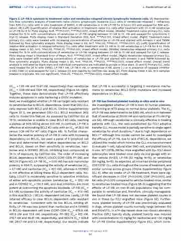
Figure 2. LP-118 is cytotoxic to treatment-naïve and venetoclax-relapsed chronic lymphocytic leukemia cells. (A) Representa- tive flow cytometry analysis of treatment-naïve chronic lymphocytic leukemia (CLL) cells or venetoclax-relapsed / -refractory (Ven R/R) CLL cells with in vitro treatment of DMSO, 4 nM venetoclax or 4 nM LP-118 for 8 hours (hr) and assayed for BAK acti- vation. (B) (Left) Quantification of relative BAK activation in treatment-naïve CLL cells after treatment with 0.1-4 nM venetoclax or LP-118 for 8 hr. Plots display, N=8. ***P<0.001, ****P≤0.0001, mixed effect model. (Middle) Treatment-naïve primary CLL cells treated for 12 hr with concentrations of venetoclax or LP-118 ranging between 0.1 nM to 10 nM and assayed for cytochrome C (Cyt C) release. Plots display mean ± Standard Deviation (SD), N=4. ***P≤0.001, mixed effect model. (Right) Treatment-naïve primary CLL cells were treated with increasing concentrations of venetoclax or LP-118 and stained with Annexin V and TMRM followed by flow cytometry analysis. Plots display mean ± SD, N=7. *P≤0.05, **P≤0.01, ***P≤0.0001. (C) (Left) Quantification of relative BAK activation in relapse/refractory CLL cells after treatment with 0.1 nM to 10 nM venetoclax or LP-118 for 8 hr. Plots display mean ± SD, N=5. *P≤0.05, **P≤0.01, ***P≤0.001, mixed effect model. (Middle) Venetoclax-relapsed primary CLL cells treated for 15 hr with concentrations of venetoclax or LP-118 ranging between 0.1 nM to 10 nM and assayed for Cyt C release. Plots display mean ± Standard Error of Mean, N=3. ****P≤0.0001, mixed effect model. (Right) Venetoclax-relapsed primary CLL cells were treated with increasing concentrations of venetoclax or LP-118 and stained with Annexin V and TMRM followed by flow cytometry analysis. Plots display mean ± SD, N=4. *P≤0.05, **P≤0.01, ****P≤0.0001, mixed effect model. Overall trend analysis is indicated by black stars and individual comparisons are indicated by purple or blue stars. (D and E) Primary CLL cells were treated for 24 hr with DMSO, LP-118 1 nM, LP-118 4 nM, or venetoclax 4 nM, alone (-) or in presence of caspase inhibitor Z-VAD-FMK (+) and assayed for Cyt C release (D) and viability by CellTiter-Glo assay (E). Plots display mean ± SD, N=3 samples treated in triplicate. NS: not significant, *P≤0.05, **P≤0.01, ****P≤0.0001, mixed effect model.
(IC50s = 1.93 nM and 15.67 nM, respectively) than venetoclax (IC50s = 3.58 nM and 1324 nM, respectively) (Figure 4A-right). Together, these data demonstrate that LP-118 effectively induces apoptosis in cells harboring a G101V mutation. Next, we investigated whether LP-118 can target cells resistant to venetoclax due to BCLXL dependence. Given that OSU-CLL cells express both BCL2 and BCLXL (Online Supplementary Figure S1 I), we generated OSU-CLL BCL2 knockout (KO) cells to model this feature. As assessed by CellTiter-Glo at 72 hr, venetoclax is unable to clear BCL2 KO cells, reaching a plateau at 50% killing above 200 nM. In contrast, LP-118 is effective at eliminating BCL2 KO cells with an IC50 of 29.5 nM versus 0.26 nM for WT cells (Figure 4B). To further charac- terize the relative potency of LP-118 in cells with increasing dependency on BCLXL, we used a panel of 5 lymphoid cell lines and determined their relative dependencies on BCL2 and BCLXL based on their sensitivity to venetoclax, navi- toclax and A-1331852 (BCLXL inhibiting tool compound) at 24 hr of exposure, by CellTiter-Glo. The order of increasing BCLXL dependency is: RS4;11, LOUCY, CCRF-CEM, PF-382, and MOLT4 (Figure 4C). LP-118 (IC50 = 0.05 nM) has sub-nanomolar potency in RS4;11, surpassing the activity of venetoclax (IC50 = 4.7 nM) and navitoclax (IC50 = 42.1 nM), whereas A-1331852 is not effective at killing these BCL2-dependent cells. No- tably, LOUCY is moderately sensitive to selective inhibitors of either anti-apoptotic protein (venetoclax IC50 = 125.1 nM and A-1331852 IC50= 130.3 nM), and dual inhibitors are most potent at overcoming the apoptosis blockade. LP-118 (IC50 = 5.5 nM) surpasses the activity of navitoclax (IC50 = 47.4 nM) in this dual BCL2- / BCLXL-dependent cell line. Lastly, LP-118 retained efficacy to clear BCLXL-dependent cells resistant to venetoclax. Consistent with its low BCLXL binding af- finity, LP-118 was less potent than navitoclax and A-1331852 in BCLXL-dependent cell lines (CCRF-CEM IC50s= 979.9 nM, 491.9 nM and 13.9 nM, respectively; PF-382 IC50= 812 nM, 378.7 nM and 46.61 nM, respectively; and MOLT4 IC50= 626.8 nM, 290.7 nM and 5.5 nM, respectively). Our results indicate
that LP-118 is successful in targeting 2 resistance mecha- nisms to venetoclax: BCL2 G101V mutations and increased dependence on BCLXL.
LP-118 has limited platelet toxicity in vitro and in vivo
We investigated whether LP-118 is toxic to human platelets, performing an MTS assay on normal donor platelets. The IC50 of LP-118 was determined to be 6360 nM, which is in-between that of venetoclax at 25000 nM and navitoclax at 170 nM (Fig- ure 5A). Although venetoclax is clinically effective in treating patients with CLL, the well-established Eμ-TCL1 adoptive transfer CLL mouse model is partially sensitive to single-agent venetoclax for short durations,24 due to high dependence on MCL1.25 Although this model cannot be used to investigate the efficacy of LP-118, due to lack of BCLXL dependence, we utilized this model which mimics the CLL microenvironment to evaluate T-cell, natural killer (NK)-cell, and platelet toxicity in vivo. WT C57BL/6NTac mice engrafted with Eμ-TCL1 donor splenocytes were treated once daily via oral gavage with ei- ther vehicle (N=20), LP-118 (50 mg/kg, N=15), or venetoclax (50 mg/kg, N=15). As expected, all mice had similar peripheral CD5+CD19+ CLL cells throughout the course of treatment, and similar survival across groups (Online Supplementary Figure S1J, K). After six weeks of LP-118 treatment, there were sig- nificant decreases in CD4+ (P=0.0434), CD8+ (P<0.0001), and NK cells (P<0.001) compared to vehicle-treated mice, similar to venetoclax-treated mice (Figure 5B-D) suggesting that the effects of LP-118 on non-B-cell populations may be com- parable to venetoclax and, therefore, clinically manageable. We found that LP-118 does not decrease platelet counts in vivo in these Eμ-TCL1 engrafted mice (Figure 5E). Further- more, platelet toxicity of LP-118 was preclinically evaluated in dogs, whose platelet counts normally range between 150 and 500x109/L. In a 14-day repeat-dose non-Good Laboratory Practice (GPL) toxicity study, platelet toxicity was induced with concentrations >3 mg/kg for navitoclax and >30 mg/kg for LP-118 (Online Supplementary Figure S1L), which corre-
Haematologica | 110 January 2025
84
