
Page 46 - Haematologica Vol. 110 - January 2025
P. 46
PERSPECTIVE ARTICLE
G. Semenzato et al.
appropriate diagnostic interpretation that distinguishes T-CUS from its mimickers, in particular T-LGLL and other borderline T-cell malignancies.
T-cell clones in healthy subjects
During a primary immune response against antigens, T-cell expansions are usually polyclonal, but oligoclonal or strictly monoclonal populations might develop, which can some- times make the differential diagnosis from T-cell malig- nancies challenging. The emergence of T-cell clones after cell activation is a normal occurrence under physiological conditions, with these T-cell expansions typically generated as a part of a reactive immune response. The detection of a clonal expansion can sometimes represent the extreme larger-than-expected physiological proliferation of cytotoxic clonotypes central to adaptive immunity, which does not necessarily imply neoplasia.1,13
Following the clearance of the relevant stimuli, proliferating cells undergo activation-induced cell death. This process helps to maintain immune homeostasis by eliminating ex- cessive or unnecessary immune responses. Reactive T-cell expansions are transient, usually self-limited, and typically observed in association with well-defined triggering events, primarily viral infections, but not only. Since reactive T-cell expansions typically resolve once the infection is cleared, the persistence of a small abnormal population over time (usually 6 months) is required before considering the pos- sibility of malignancy.
Given the essentially indistinguishable morphology of leu- kemic and reactive large granular lymphocytes (LGL), proof of clonality is mandatory. This confirmation is primarily based on the unique structure of the T-cell receptor (TCR) which is thought to carry the fine specificity for the an- tigen in its hypervariable complementarity-determining region 3 (CDR3). Clonality of T cells is easily detectable by conventional molecular technologies based on polymerase
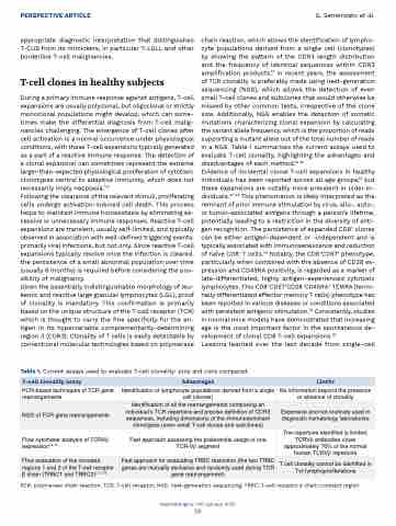
chain reaction, which allows the identification of lympho- cyte populations derived from a single cell (clonotypes) by showing the pattern of the CDR3 length distribution and the frequency of identical sequences within CDR3 amplification products.14 In recent years, the assessment of TCR clonality is preferably made using next-generation sequencing (NGS), which allows the detection of even small T-cell clones and subclones that would otherwise be missed by other common tests, irrespective of the clone size. Additionally, NGS enables the detection of somatic mutations characterizing clonal expansion by calculating the variant allele frequency, which is the proportion of reads supporting a mutant allele out of the total number of reads in a NGS. Table 1 summarizes the current assays used to evaluate T-cell clonality, highlighting the advantages and disadvantages of each method.14-18
Evidence of incidental clonal T-cell expansions in healthy individuals has been reported across all age groups,19 but these expansions are notably more prevalent in older in- dividuals.20-22 This phenomenon is likely interpreted as the remnant of prior immune stimulation by virus, allo-, auto-, or tumor-associated antigens through a person’s lifetime, potentially leading to a restriction in the diversity of anti- gen recognition. The persistence of expanded CD8+ clones can be either antigen-dependent or -independent and is typically associated with immunosenescence and reduction of naïve CD8+ T cells.23 Notably, the CD8+CD57+ phenotype, particularly when combined with the absence of CD28 ex- pression and CD45RA positivity, is regarded as a marker of late-differentiated, highly antigen-experienced cytotoxic lymphocytes. This CD8+CD57+CD28–CD45RA+ TEMRA (termi- nally differentiated effector memory T cells) phenotype has been reported in various diseases or conditions associated with persistent antigenic stimulation.24 Consistently, studies in normal mice models have demonstrated that increasing age is the most important factor in the spontaneous de- velopment of clonal CD8 T-cell expansions.25
Lessons learned over the last decade from single-cell
Table 1. Current assays used to evaluate T-cell clonality: pros and cons compared.
T-cell clonality assay
Advantages
Limits
PCR-based techniques of TCR gene rearrangements
Identification of lymphocyte populations derived from a single cell (clones)
No information beyond the presence or absence of clonality
NGS of TCR gene rearrangements
Identification of all the rearrangements composing an individual’s TCR repertoire and precise definition of CDR3 sequences, including dimensions of the immunodominant clonotypes (even small T-cell clones and subclones)
Expensive and not routinely used in diagnostic hematology laboratories
Flow cytometer analysis of TCRVβ expression15,16
Fast approach assessing the preferential usage of one TCR-Vβ segment
The repertoire identified is limited, TCRVβ antibodies cover approximately 70% of the normal human TCRVβ repertoire
Flow evaluation of the constant regions 1 and 2 of the T-cell receptor β chain (TRBC1 and TRBC2)7,17,18
Fast approach for evaluating TRBC restriction (the two TRBC genes are mutually exclusive and randomly used during TCR gene rearrangement)
T cell clonality cannot be identified in Tgd lymphoproliferations
PCR: polymerase chain reaction; TCR: T-cell receptor; NGS: next-generation sequencing; TRBC: T-cell receptor β chain constant region.
Haematologica | 110 January 2025
38
