
Page 24 - Haematologica Vol. 110 - January 2025
P. 24
EDITORIAL
Challenges in defining the immune microenvironment in T-cell lymphoma
Ahmet Dogan and Mikhail Roshal
Department of Pathology and Laboratory Medicine, Memorial Sloan Kettering Cancer Center, New York, NY, USA
In this issue of Haematologica, Stephan et al. report a de- tailed single-cell immune profiling of T cells in peripheral T-cell lymphomas (TCL).1 Immuno-oncology approaches including immune checkpoint blockade,2 chimeric antigen receptor (CAR) T cells,3 and bi-specific therapeutic antibod- ies4 have been effectively employed in the management of B-cell lineage lymphomas (BCL). For many BCL subtypes, these therapies have altered the standard of care. The success of these treatments is partly attributed to a deep understanding of the underlying immunobiology of B-cell lineage lymphomas.5-8 Although there is growing interest in applying similar therapeutic strategies to TCL, progress has been limited. This is due in part to the rarity and the biological complexity of these disorders, but the primary
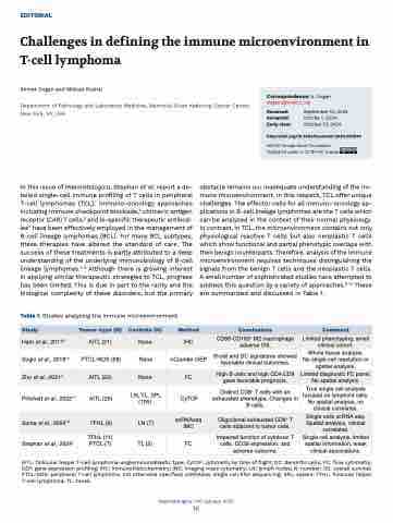
Table 1. Studies analyzing the immune microenvironment.
obstacle remains our inadequate understanding of the im- mune microenvironment. In this respect, TCL offer unique challenges. The effector cells for all immuno-oncology ap- plications in B-cell lineage lymphomas are the T cells which can be analyzed in the context of their normal physiology. In contrast, in TCL, the microenvironment contains not only physiological reactive T cells but also neoplastic T cells which show functional and partial phenotypic overlaps with their benign counterparts. Therefore, analysis of the immune microenvironment requires techniques distinguishing the signals from the benign T cells and the neoplastic T cells. A small number of sophisticated studies have attempted to address this question by a variety of approaches.9-13 These are summarized and discussed in Table 1.
Study
Tumor-type (N)
Controls (N)
Method
Conclusions
Comment
Ham et al., 20179
AITL (21)
None
IHC
CD68-CD163+ M2 macrophage adverse OS.
Limited phenotyping, small clinical cohort.
Sugio et al., 201810
PTCL-NOS (68)
None
nCounter GEP
B-cell and DC signatures showed favorable clinical outcomes.
Whole tissue analysis. No single cell resolution or spatial analysis.
Zhu et al., 202111
AITL (50)
None
FC
High B cells and high CD4:CD8 gave favorable prognosis.
Limited diagnostic FC panel. No spatial analysis.
Pritchett et al., 202212
AITL (25)
LN, TL, SPL (105)
CyTOF
Distinct CD8+ T cells with an exhausted phenotype. Changes in B cells.
True single cell analysis focused on lymphoid cells. No spatial analysis, no clinical correlates.
Suma et al., 202413
TFHL (9)
LN (7)
scRNAseq IMC
Oligoclonal exhausted CD8+ T cells adjacent to tumor cells.
Single cells scRNA seq. Spatial analysis, clinical correlates.
Stephan et al., 20241
TFHL (11) PTCL (7)
TL (5)
FC
Impaired function of cytotoxic T cells, CD39 expression, and adverse outcome.
Single cell analysis, limited spatial information, weak clinical associations.
AITL: follicular helper T-cell lymphoma-angioimmunoblastic type; CyTOF: cytometry by time of flight; DC: dendritic cells; FC: flow cytometry; GEP: gene expression profiling; IHC: immunohistochemistry; IMC: imaging mass cytometry; LN: lymph nodes; N: number; OS: overall survival; PTCL-NOS: peripheral T-cell lymphoma, not otherwise specified; scRNAseq: single cell RNA sequencing; SPL: spleen; TFHL: follicular helper T-cell lymphoma; TL: tonsil.
Haematologica | 110 January 2025
16
Correspondence: A. Dogan dogana@mskcc.org
Received: Accepted: Early view:
September 10, 2024. Octobe 1, 2024. October 10, 2024.
https://doi.org/10.3324/haematol.2024.285836
©2025 Ferrata Storti Foundation Published under a CC BY-NC license
