


Page 145 - Haematologica Vol. 110 - January 2025
P. 145
ARTICLE - Immune microenvironment in nodal PTCL
P. Stephan et al.
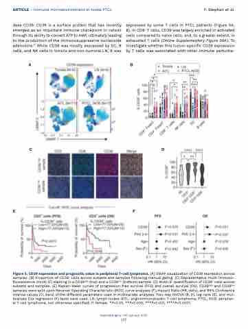
dase CD39. CD39 is a surface protein that has recently emerged as an important immune checkpoint in cancer through its ability to convert ATP to AMP, ultimately leading to the production of the immunosuppressive nucleoside adenosine.21 While CD39 was mostly expressed by DC, B cells, and NK cells in tonsils and non-tumoral LN, it was
expressed by some T cells in PTCL patients (Figure 5A, B). In CD8+ T cells, CD39 was largely enriched in activated cells compared to naïve cells, and, to a greater extent, in exhausted T cells (Online Supplementary Figure S8A). To investigate whether this tumor-specific CD39 expression by T cells was associated with other immune perturba-
 AB
CD
EF
Figure 5. CD39 expression and prognostic value in peripheral T-cell lymphoma. (A) UMAP visualization of CD39 expression across samples. (B) Proportion of CD39+ cells across subsets and samples following manual gating. (C) Representative multi-immuno- fluorescence (multi-IF) staining in a CD39high (top) and a CD39low (bottom) sample. (D) Multi-IF quantification of CD39+ cells across subsets and samples. (E) Kaplan-Meier curves of progression-free survival (PFS) and overall survival (OS). CD39high and CD39low samples were split upon Receiver Operating Characteristic (ROC) curve analyses (F), Hazard Ratio (HR, dots), and 95% Confidence Interval values (CI, bars) of the different parameters used in multivariate analyses. Two-way ANOVA (B, D), log-rank (E), and mul- tivariate Cox regression (F) tests were used. LN: lymph nodes; AITL: angioimmunoblastic T-cell lymphoma; PTCL, NOS: peripher- al T-cell lymphoma, not otherwise specified; F: female. *P<0.05, **P<0.005, ***P<0.001, ****P<0.0001.
Haematologica | 110 January 2025
137


