
Page 63 - Haematologica-5
P. 63
staining). RONi treatment did not affect mesangial expan- sion in SCD mice (Figure 8A and B, H&E and Online Supplementary Figure S6, PAS staining). However, RONi administration in SCD mice significantly reduced capillary expression of vWF (Figure 8C and G and Online Supplementary Figure S5, immunostaining, red), and Intercellular Adhesion Molecule 1 (ICAM1) (Figure 8D and E and Online Supplementary Figure S5, immunostain- ing, red), which are the markers of endothelial injury.36,37 Thus, inhibiton of RON receptor in SCD mice significant- ly ameliorated development of endothelial and glomerular injury.
Discussion
Renal disease in SCD patients includes a variety of glomerular and tubular complications, but the mechanism of their development is not fully understood. The most important finding in our study is that MSP1 and its recep- tor RON kinase may play a role in the activation of renal endothelium and development of glomerular pathology in SCD mice. Moreover, treatment of mice with RONi, an inhibitor of RON kinase, significantly ameliorated glomerular hypertrophy, capillary dilation and congestion, and endothelial injury. These findings reveal a previously unknown mechanism which contributes to glomerular endothelial injury in SCD.
Sickle cell disease mice spontaneously develop FSGS that may be directly associated with RBC sickling and chronic hemolysis. The focal nature of glomerulosclerosis in SCD mice and SCD patients apparently excludes the effect of global factors, such as hypoxia, cell-free heme, iron, and other circulating factors that would lead to a global and not focal glomerulosclerosis with involvement of only 50% of glomeruli in SCD mice. Thus, locally-pro- duced factor(s) are more likely to contribute to the devel- opment of FSGS.
In agreement with a previous report,21 we demonstrate here that macrophage infiltration in renal glomeruli was increased in SCD mice. Sickling and adhesion of RBCs, and accumulation of RBC lysate products within the kid- ney might stimulate renal infiltration of monocyte-derived macrophages. Monocyte-derived renal macrophages are present in all forms of kidney disease with inflammation, and renal capillary macrophage infiltration is a character- istic pathology of FSGS.38 In many human biopsy studies, number of glomerular or interstitial macrophages correlate with poor outcomes, suggesting their possible role in the disease progression.39,40 However, the role of infiltrating macrophages in the progression of renal disease is not well understood. Phagocytosis of senescent sickled RBCs, RBC exosomes and endocytosis of cell-free hemoglobin increases inflammatory response in the cultured human monocytes/macrophages.12 Activation of proteases is a universal inflammatory response in macrophages.41 We demonstrate here that products of RBC hemolysis signifi- cantly increased expression of macrophage membrane- bound protease, MT-SP1 in cultured human macrophages. Meta-analysis data also showed that hypoxia and mono- cyte differentiation increased MT-SP1 expression in pri- mary human macrophages. MT-SP1 is a type II membrane serine protease that plays important roles in cell migration and tumor cell metastasis.42
MT-SP1 is one of the proteases that activate circulating
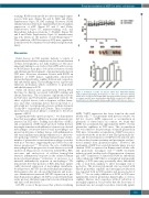
Figure 7. Treatment of sickle cell disease (SCD) mice with RON inhibitor
haematologica | 2018; 103(5)
Renal accumulation of MSP1 in sickle cell disease
A
B
C
P=0.04
reduces kidney hypertrophy. (A) Treatment of
(RONi) does not affect body weight (BW). (B) Representative picture of kidneys of controls and SCD mice treated either with (RONi) or vehicle (2% DMSO). (C) Quantification of kidney weight. Kidney weights (KW) to BW ratios are shown.
SCD mice
with RON inhibitor
MSP1.15 MSP1 expression has been found in the renal tubular cells.43,44 In agreement with previous studies, we did not observe MSP1 expression or accumulation in glomeruli of control mice. In contrast, we found that MSP1 was accumulated in approximately 46±8% of renal glomerular capillaries in SCD mice. This accumulation rate was similar to the percentage of injured glomeruli in SCD mice. Glomerular accumulation of MSP1 was previ- ously shown in the rat model of anti-Thy1 glomerular dis- ease, and the neutralization of MSP1 by the injected anti- bodies reduced serum creatinine and proteinuria, and pro- tected animals from glomerular injury.17 However, the mechanism of MSP1-associated glomerular injury was not clarified. RON is expressed in human renal tubular cells and glomerular mesangial cells.43,44 MSP1 treatment induces growth, motility and collagen invasion of mesan- gial cells.43 Expression of functional RON in endothelial cells is unknown. MSP1 that is accumulated in glomerular capillary of SCD mice may potentially affect endothelial cells, or leak from capillary to affect mesangial cells or podocytes. The increased glomeruli size associated with dilated glomerular capillary and mesangial proliferation was reported both in SCD patients and mouse model of SCD.20,21,45 In our study, inhibition of RON in SCD mice significantly reduced glomerular hypertrophy, as well as capillary dilation and congestion without reduction of mesangial expansion. It is possible that the short time of treatment was not enough to produce statistically signifi-
795
