
Page 61 - Haematologica-5
P. 61
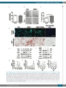
Figure 5. MSP1 treatment of cultured human glomerular endothelial cell line (HGEC) induces cell motility and vWF expression, F–actin re-organization, and RON kinase signaling in HGEC. (A) Cell growth measured by MTT assay. Five wells are used for either control or treatment with 1 μM of recombinant MSP1 in each exper- iment. Results are representative of three independent experiments. (B) Representative picture of wound migration assay of control cells and cells treated with 1 μM of MSP1. Bar size 300 μm. (C) Quantification of wound migration assay. Three wells are used per treatment in each experiment. Results are representative of three independent experiments. (D) Immunostaining of F-actin (green) and vWF (red) in control and treated MSP1 (1 μM) treated cells with or without RONi (200 nM). DAPI (for F-actin) and Hematoxylin (for vWF) were used for nuclear staining. Non-specific primary antibodies were used for negative control. Bar size 40 μm for F-actin and 100 μm for vWF. (E and F) Western blots of phosphorylated and non-phosphorylated forms of ERK and AKT kinases in HGEC. (E) Cells were treated with MSP1 (1 μM) and collected at different time points after treatment. (F) Cells were treated with MSP1 (1 μM) with or without RON inhibitor (RONi, 200 nM) for 30 min. (G-J) Quantification of pERK and pAKT on Western blot. For quantification graphs, mean and SD are shown. *P<0.05. Results are representative of three inde- pendent experiments. β-actin used for loading normalization.
Renal accumulation of MSP1 in sickle cell disease
ABC
D
EF
GHIJ
P=0.003
haematologica | 2018; 103(5)
793
