
Page 51 - Haematologica-5
P. 51
Stem cell mobilization in sickle cell patients
for P1, P2 and P3, respectively) were high in all three patients despite a limited collection efficiency (Table 2). This enabled the cryopreservation of 3x10⁶ unselected CD34+ cells/kg as a back-up for the upcoming gene thera- py trial (used in the case of absence of engraftment) and the immunoselection and further analyses of mobilized CD34+ cells as detailed below. Following CD34+ selection, CD34+ cell purity was in the normal range (Table 2). CD34+ recovery was high in P1 and P2 (82% and 92%, respectively) and lower in P3 (31%).
Characterization of mobilized hematopoietic stem and progenitor cells
To determine the hematopoietic differentiation capacity and self-renewal potential of the mobilized CD34+ cells, we performed a number of phenotypic, transcriptomic and functional analyses.
Hematopoietic stem cells (HSC) and their immediate progeny (multipotent progenitors) within the CD34+ sub- set are negative for lineage, CD38, and CD45RA markers and positive for CD13323-25 and can, therefore, be detected using flow cytometry. We compared the numbers of HSC and multipotent progenitors among mobilized SCD CD34+ cells with the values for (i) the BM of healthy donors and SCD patients, and (ii) samples from healthy donors mobilized with either Filgrastim or Plerixafor (Online Supplementary Table S1, Figure 1C and Online Supplementary Figure S2). We estimated the number of
HSC per 1000 CD34+ cells to be >25 in Plerixafor-mobi- lized SCD samples and <5 in all other samples, suggesting that Plerixafor mobilizes HSC with an unexpectedly high efficacy in SCD patients.
The frequency of erythroid and granulocyte/monocyte colony-forming cells was similar in Plerixafor-mobilized and BM SCD samples and in the range of that observed in Filgastrim-mobilized HSPC (Online Supplementary Figure S3A and data not shown). Upon erythroid differentiation, Plerixafor-mobilized as well as BM SCD HSPC gave rise to >90% of mature GYPA+CD36lowCD71low enucleated red blood cells (Online Supplementary Figure S3B-D).
The transcriptome of highly purified CD34+ HSPC from the different sources was analyzed using RNA-Seq. Unsupervised gene expression analysis showed that the samples clustered into two major groups, based on cell origin: the “BM” group encompassed BM samples from SCD patients and healthy donors, whereas the “mobi- lized” group encompassed Plerixafor-mobilized HSPC from SCD patients and healthy donors and Filgrastim- mobilized HSPC from healthy donors (Figure 2A).
Next, we identified differentially expressed genes among the different populations (false discovery rate <0.05, Table 3). In a comparison of samples from SCD patients and healthy donors, we observed that the genes upregulated in SCD samples are involved in inflammatory and immune responses (e.g. defense response to other organisms, type I interferon signaling pathway, cytokine
A
B
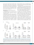
Figure 3. Plerixafor-mobilized CD34+ cells from sickle cell disease patients engraft to the same degree as Filgrastim-mobilized CD34+ cells from healthy donors in NSG mice. NSG mice were sacrificed 3 to 4 months after the injection of SCD (SCD Plerixafor, n=3) or HD (HD Filgrastim, n=2) CD34+ cells. (A) Bone marrow cells and (B) splenocytes were isolated, stained and analyzed by flow cytometry. The chimerism (defined as % human CD45+cells/total CD45+cells) and the numbers of human B lymphocytes (CD19+IgM+), granulocytes (CD11b+CD15+), and monocytes (CD11b+CD14+) were evaluated in each group of mice (red circles and red triangles SCD Pler1; blue circles and blue triangles SCD Pler2; green circles and green triangles SCD Pler3; the two HD Filg control are represented by gray squares/gray dia- mond and black squares/black diamonds, respectively). Each dot represents an individual mouse. HD: healthy donor; SCD: sickle cell disease; Pler: Plerixafor; Filg: Filgrastim.
haematologica | 2018; 103(5)
783
