
Page 43 - Haematologica-5
P. 43
B
++ berofcellsforTF mono,aβ2 PMN,and
haematologica | 2018; 103(5)
Safety and efficacy of plerixafor in SCD patients
A
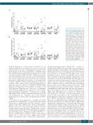
Figure 4. Fold changes in the percent- age of cells expressing activation mark- ers, according to plerixafor dose. (A) Other than % TF+ monocytes, median fold changes in % cells were ≤1.1. (B) Median fold changes in absolute num-
with the findings of a recent study by Tisdale et al., in which mobilization was effective in seven of seven SCD subjects (100%) at a dose of 240 μg/kg.4 It should be noted that patients in the National Institutes of Health study were off hydroxyurea and had been transfused for at least 2 months to achieve a HbS <20-30%4,32 while in our study, ten of the 15 patients were on stable doses of hydroxyurea (for at least 1 year) and only one patient was on chronic transfusion. Although hydroxyurea, a ribonucleotide reductase inhibitor, causes myelosuppression and was recently found to reduce CD34 counts in peripheral blood and bone marrow,33 there is no definitive evidence that hydroxyurea negatively affects numbers or quality of cell cycle-quiescent hematopoietic stem cells or immature bone marrow progenitors as opposed to more mature myeloid-erythroid progenitors.33-37 Indeed, in our study, although we did not achieve consistent efficiency in CD34 cell mobilization, no correlation was found between hydroxyurea use, and absolute or fold increases in CD34+ cells/μL.
We observed wide inter-donor variability in CD34 mobilization with plerixafor, as previously reported in normal donors (CD34 peaks between 4-157/μL )13 and in patients with SCD (CD34 peaks between 50-200/μL).4 However, we also observed a strong correlation between baseline CD34+ and peak CD34+ concentrations, as previ- ously reported with both G-CSF and plerixafor mobiliza- tion in healthy donors (Kendall tau=0.68, P=0.0006).13,30,31 Factors contributing to baseline CD34 count remain unclear, but our data and others’ suggest that baseline CD34+ concentration may be affected by hydroxyurea-
aMac-1+ PMN were closer to 2 than 1. Fold changes are ratios of 12 h to base- line values. Horizontal lines represent medians. Filled shapes represent hydroxyurea-treated patients, open shapes represent patients not treated with hydroxyurea. Gray arrows point to the values for the two SAE patients who had serious adverse events. TF+ mono: tissue-factor-positive monocytes; E-Sel+ PMN: E-selectin-positive neutrophils; aβ2+ PMN: activated β2 integrin-positive neutrophils, aMac-1+ PMN: activated Mac-1-positive neutrophils; L-Selneg PMN: L-selectin-negative neutrophils; PMA: platelet-monocyte aggregates; PNA: platelet-neutrophil aggregates.
related myelosuppression.24,33 Patient #8, a subject re- enrolled in the study, was particularly instructive regard- ing this hypothesis. This patient was clinically stable on hydroxyurea at a dose of 27 mg/kg and was enrolled twice at an interval of 13 months. At the time of his second treatment, however, he had a markedly lower baseline ANC (1900/μL down from 6300/μL) and platelet count (217,000/μL down from 400,000/μL), probably related to oscillatory non-toxic hematopoiesis seen in SCD with chronic and dose-intensive treatment with hydroxyurea (ANC oscillations between 2,000-6,000/μL as determined from review of his clinical laboratory records).38 This myelosuppression was associated with a baseline CD34 concentration of 0/μL rather than 1/μL, possibly contribut- ing to the relatively low 12 h CD34+ concentration of 10/μL at the 240 μg/kg dose as compared to 27/μL at the 160 μg/kg dose. In brief, because hydroxyurea can decrease ANC and platelet count,39 hydroxyurea-related myelosuppression may have contributed to the relatively poor CD34+ mobilization obtained in this cohort. However, avoiding hydroxyurea withdrawal might lower the risk of pain crises;24 we, therefore, plan to explore tim- ing plerixafor administration to the peak rather than nadir of hydroxyurea-related oscillatory hematopoiesis. Finally with regards to hydroxyurea therapy, data from the six patients in whom we enumerated CD34+CD38– cells sug- gest that hydroxyurea may not adversely affect HSC, given that all patients except one (patient 10) were on hydroxyurea and a median 3-fold increase at 12 h was observed. Only 0.2-2.8% of CD34+ cells were CD38-neg- ative, but this may be consistent with plerixafor’s effect in
775
