
Page 40 - Haematologica-5
P. 40
F. Boulad et al.
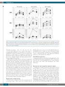
Figure 1. Peripheral blood white blood cell, absolute neutrophil, and CD34 cell counts. There is a trend of increasing white blood cell count (WBC, P=0.05) and absolute neutrophil count (ANC, P=0.03), but not CD34 concentration (P=0.65) with increasing dose of plerixafor. The graphs show the peripheral blood WBC, ANC, and CD34 concentrations of 15 patients with SCD treated whith 80 (circles), 160 (squares) and 240 (triangles) μg/Kg of plerixafor prior to administration of plerixafor (PRE), 6-12 h and 20-24 h after plerixafor. Patients on hydroxyurea are represented by filled circles, squares and triangles, patients off hydroxyurea are represented by open circles, squares and triangles.
772
Probability of escalation: 0.91 0.71 0.49 0.31 0.17 0.08 For the efficacy endpoint, the dose escalation will continue to 480 μg/kg unless all patients at a preceding dose level achieve a peripheral blood CD34+ concentration of at least 30 cells/μL. In the present dose-escalation phase, no leukapheresis is performed. If and when the efficacy endpoint is safely reached, the study will proceed to a leukapheresis phase (including preclinical transduc-
Dickinson Biosciences, San Jose, CA, USA) and FACS Diva soft- ware (BD Biosciences). Samples are stained and analyzed within 2-12 h of collection using a modification of the International Society of Hematotherapy and Graft Engineering (ISHAGE) method (see Online Supplementary Methods).
CD34+CD38– enumeration
Mononuclear cells are isolated from 2 mL peripheral blood by Ficoll-Hypaque Plus density centrifugation. CD34+ cells are puri- fied by positive selection (MidiMACSTM LS Columns, Miltenyi) and stained with CD34 (BD PharMingen) and CD38 (Invitrogen).
Research cell and coagulation activation studies
Peripheral blood samples drawn at baseline and after 12 h are stained within 1 h of collection for activation markers relevant to sickle vaso-occlusion12,25-28 and assessed by flow cytometry (BD FACSCantoTM). For CD16b+ (1D3, Beckman Coulter) neu- trophils: activated β2 integrin (clone 24, abcam), activated Mac- 1 (CBRM1/5, eBioscience), E-selectin-Fc chimera (724-ES, R&D Systems), L-selectin (DREG-56, eBioscience), Mac-1/CD11b (ICRF44, BD Pharmingen), and LFA-1/CD11a (HI111, BD Pharmingen). For CD14+ (M5E2, BD Pharmingen) monocytes: tissue factor (HTF-1, BD Pharmingen). For CD41+ (HIP2, BD Pharmingen) platelets: CD16b (1D3, Beckman Coulter) and CD14 (M5E2, BD Pharmingen). The percentages of positive cells and median fluorescent intensity (MFI) are assessed for each
tion and editing) in three patients.
Eligible subjects are adults with SS or Sβ0 disease, normal renal
and liver function, hemoglobin concentration ≥6 g/dL, WBC count ≥3,000/μL, absolute neutrophil count (ANC) ≥1,500/μL, and platelet count of ≥150,000/μL. Eligible subjects are admitted to the Clinical Research Center at Weill Cornell Medical College. A sin- gle subcutaneous injection of plerixafor (Sanofi-Genzyme) is administered in the evening between 8-9 pm. The protocol calls for peripheral blood sampling at three time points (baseline, 0-2 h prior to plerixafor; peak between 6-12 h after the plerixafor dose; at the presumed return to baseline between 20-24 h after the dose): for reasons of feasibility and patient comfort issues, the peak sample was consistently drawn at a mean of 12 ± 1 h after plerixafor administration and the return to baseline sample at a mean of 20 ± 0.29 h after the dose. Since patients have pre-existing anemia, for reasons of safety no more than a total of 105 mL of blood is drawn at all three time points combined.
Peripheral blood CD34 testing
Flow cytometric evaluation of the collected peripheral blood is performed using a FACS Canto flow cytometer (Becton
haematologica | 2018; 103(5)
