
Page 172 - Haematologica-5
P. 172
P.H. Mangin et al.
activation of GPVI by platelet-bound fibrinogen partici- pates in platelet aggregation we performed an in vitro flow adhesion assay under conditions that prevent activation of GPVI by collagen and by fibrin. To achieve this, we gener- ated a platelet aggregate over type I fibrillar collagen using hirudin-treated blood to prevent formation of fibrin. We
A
then perfused additional blood from the same donor over the aggregate at a wall shear rate of 300 s-1 in the presence or absence of the Fab fragment of the GPVI blocking mon- oclonal antibody 9O12. As expected, we were unable to detect the presence of fibrin in the aggregate using a spe- cific antibody (data not shown). The aggregate continued to
904
B
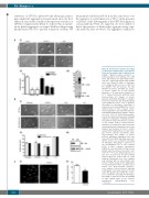
Figure 4. Syk promotes platelet spreading on fibrinogen downstream of glycoprotein VI but not in integrin aIIbβ3 outside-in sig- naling. Washed human platelets, or washed platelets from wild-type mice (WT mice), Syk chimera mice (Syk chimera mice) and mice expressing human GPVI (hGPVI mice) were allowed to adhere to fibrinogen or to collagen in the presence of either the Src inhibitor PP2 (20 μmol/L), the Syk inhibitor (5 μmol/L), thrombin (0.1 U/mL), or vehicle control, for 30 min (human platelets) or 45 min (mouse platelets) at 37°C followed by fixation with PFA. (A)(i). Representative DIC images of human platelets adhering to fibrinogen or collagen. Scale bars represent 5 μm. (A)(ii). Bar graph representing the surface area of platelets spreading on fibrinogen. Spreading is expressed as the mean±SEM in five or more random fields, in three separate experi- ments. Significance was determined by one- way ANOVA and Bonferroni multiple compar- ison test: *P<0.01. (A)(iii) Representative western blot from human platelets following adhesion to fibrinogen. Following immuno- precipitation of Syk, proteins were separat- ed by sodium dodecyl sulfate-polyacry- lamide gel electrophoresis and western blot for phosphotyrosine. Membranes were then stripped and reprobed for Syk to confirm equal protein loading. Blots are representa- tive of three separate experiments. (B)(i). Representative DIC images of mouse platelets adhering to fibrinogen. Scale bars represent 5 μm. (B)(ii). Bar graph represent- ing the surface area of platelets spreading on fibrinogen. Spreading is expressed as the mean±SEM in five or more random fields, in three separate experiments. Significance was determined using one-way ANOVA, with the Bonferroni post-hoc test: *P<0.05, **P<0.001. (B)(iii). Expression of Syk was measured by western blotting platelet whole-cell lysates with an a-Syk antibody. Membranes were then stripped and reprobed with an anti-a-tubulin anti- body to confirm equal protein loading. Blots are representative of three separate experi- ments. (C)(i). Representative epifluores- cence images of mouse platelets adhering to fibrinogen. Scale bars represent 10 μm. (C)(ii). Bar graph representing the surface area of platelets spreading on fibrinogen. Spreading is expressed as the mean±SEM in eight random fields, in five separate experiments (Mann-Whitney test, **P<0.01).
C
haematologica | 2018; 103(5)
