
Page 171 - Haematologica-5
P. 171
B
haematologica | 2018; 103(5)
Fibrinogen, GPVI and platelet activation
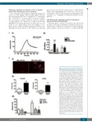
Fibrinogen stimulates an increase of Ca2+ in human glycoprotein VI-transgenic mouse platelets
The observation that platelets expressing human but not mouse GPVI undergo full spreading suggests that sig- nals from human GPVI are of significance. To investigate this, a dual-dye Ca2+ assay was used to monitor cytoplas- mic Ca2+ levels as a marker of PLCγ2 activation. Analysis of single platelet Ca2+ profiles by confocal microscopy highlighted that signals generated on fibrinogen are com- posed of Ca2+ spikes (Figure 5A). The number of Ca2+ spikes in mouse platelets expressing human GPVI was sig- nificantly increased relative to the number in wild-type
A
platelets and was blocked in the presence of PRT-060318 (Figure 5Ai-ii) highlighting the critical role of Syk in Ca2+ mobilization. These results demonstrate that human GPVI stimulates Ca2+ signaling in fibrinogen-adherent mouse platelets.
Fab 9O12 blocks aggregate growth of humanized glycoprotein VI mouse platelets
Fibrinogen plays a critical role in hemostasis and arterial thrombosis through crosslinking of platelets in the grow- ing thrombus. In addition, we now show that fibrinogen induces platlet activation by GPVI. To establish whether
Figure 3. Monomeric but not dimeric GPVI binds to immobilized fibrinogen and supports cellular adhe- sion. (A)(i). IF-1 purified fibrinogen was immobilised to a COOH-V chip surface using amine coupling covalent linkage, to a level of 3,825 RU. Monomeric or dimeric GPVI was titrated over three to four orders of magnitude across the chip surface to a maximum of 1 μmol/L, generating a single binding curve (1 RU represents the binding of approximately 1 pg/mm2 protein). The graphs shown are representative of three repeats and the KD is expressed as mean ± SEM. (A)(ii). Solid-phase binding assays were per- formed in Nunc maxisorb 96-well plates coated overnight with BSA and fibrinogen (FGN). Monomeric or dimeric GPVI (100 nmol/L) was incu- bated as described. Bound GPVI was detected using HRP-coupled to an anti-6×His monoclonal antibody for monomeric GPVI or an anti-human IgG for dimer- ic GPVI. The histogram (mean ± SD) shows the results from five independent experiments. Significance was determined using a one-way ANOVA, Dunnett post-hoc test : *P<0.05. (B). RBL- 2H3 cells (3x10⁵ cells/mL; 300 μL) transduced with an empty vector (RBL-2H3 Ctrl) or with human full- length GP6 cDNA (RBL-2H3 huGPVI) were pre-incu- bated with PBS, REOPRO (20 μg/mL), and 9O12 (50 μg/mL) and allowed to adhere to immobilized fib- rinogen for 1 h at 37°C, in 5% CO2. After three gentle washing steps, cells were permeabilised with TRI- TON x100 0.2% and stained with Alexa-568 phal- loidin (1.5 U/mL). (B)(i). Representative epifluores- cence images of RBL cells adhering to immobilized fibrinogen. (B)(ii). and (B)(iii). Cells were manually counted in six to eight different experiments (*P<0.05 Mann Whitney t-test in (ii) and one-way ANOVA was followed by the Bonferroni multiple com- parison test).
903
