
Page 149 - Haematologica-5
P. 149
Nano-scale protein quantification in multiple myeloma
marked antibodies that reduce the sensitivity of detection, and fewer antibodies are available for these techniques, even when used in an indirect assay.3 Immunohistochemistry allows only semiquantitative analysis of protein expression, and requires a well-trained pathologist to interpret the results. Moreover, neither technique is able to identify non-specific antibody binding to other proteins.3
Western blotting (WB) remains the “gold standard” tech- nique for protein characterization in most laboratories. However, WB consumes large quantities of reagents, has a low throughput, and requires a great deal of time and effort involving many laborious manual processing steps. Moreover, WB only yields semiquantitative data of poor repeatability, making it a challenge to go beyond using the assay in discovery research to apply it reliably in the clin- ical setting.4–6 A further drawback is that it is not always possible to obtain the quantity of protein extract required for WB from primary cancer samples.
MM is a clear prototype of a bone marrow-infiltrating tumor for which a relatively small quantity of sample is available after the diagnostic procedure, which involves morphological evaluation, immunophenotypic characteri- zation by flow cytometry, and CD138+ plasma cell separa- tion for routine fluorescence in situ hybridization analysis. The recent development of a method based on the combi- nation of capillary nano-electrophoresis with immunoas- say (CNIA), also known as ‘simple western’, requires only very small amounts of sample to be able to measure pro- tein expression.3,7 This technical advance makes it possible to analyze the expression of 50-100 proteins in a single MM sample. Here we present the results of a pilot study using this platform in MM patients. The main goal was to quantify accurately and robustly the proteins extracted from CD138-purified MM samples frozen in RLT Plus buffer, which is commonly used as a method for RNA and DNA preservation. Additionally, we attempted to estab- lish the clinical value of this analysis using a panel of pro- teins essential to MM pathogenesis, comparing it with that of the corresponding RNA expression.
Methods
For more specific information see the Online Supplementary File.
Patients and multiple myeloma cell lines
Sixty-three samples from patients diagnosed with MM between October 2013 and November 2015 were included in the study (Online Supplementary Table S1). Forty-three had been enrolled in two Spanish Myeloma Group clinical trials: GEM2010 [bortezomib/melphalan/prednisone and lenalidomide/dexam- ethasone in a sequential or alternating manner; (n=24)] and BenVelPres [bendamustine/bortezomib/prednisone; (n=19)]. The other 20 patients were not treated as part of a clinical trial.
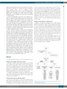
The impact of RNA and protein expression on patients’ survival was evaluated only in the group of patients that took part in the clinical trials (Online Supplementary Table S1). The scheme of the study is presented in Figure 1.
Protein extraction from RLT Plus buffer
Proteins were extracted by ice-cold acetone precipitation from RNA-column flow-through liquid. To increase the rate of protein precipitation 10 mM NaCl was added to the acetone at 80% (v/v). For technical reasons, each sample was divided into two tubes and
extracted separately. After overnight incubation at -20oC, the pro- teins were centrifuged at 13,000 x g for 30 min at 4oC, and washed twice with 70% ice-cold ethanol followed by centrifugation for a further 10 min. The protein precipitate was dried at 39oC and dis- solved with 50 μL 0.2 M NaOH for 10 min at room temperature and 4x WB sample buffer for at least 15 min at room temperature. Samples were then denatured at 95oC for 5 min, cooled to room temperature and stored in aliquots at -80oC. Before any assay, samples were heated to room temperature, then kept at 37oC for 30 min in order to re-dissolve any protein that had precipitated during freezing.
Capillary electrophoresis immunoassay
Capillary electrophoresis immunoassay or simple western analysis was performed using the WESTM machine (ProteinSimple, San Jose, CA, USA) in accordance with the manufacturer’s proto- cols. The Total Protein Assay (ProteinSimple) was used to quantify the protein concentration. In brief, 5 μL of proteins were loaded on the plate, separated by size, labeled with a biotin reagent and detected by chemiluminescence using streptavidin-horseradish peroxidase. At the end of the run, the proportion of the protein of interest in the total protein in the sample was measured, in com- parison to a standard curve previously generated using JJN3 cell line extracts of known protein concentrations.
Primary antibodies used in the study and the optimized condi- tions for each antibody are presented in Table 1. Data were ana- lyzed using CompassTM software. Each protein peak was meas- ured automatically and normalized with respect to the GAPDH median area under the peak. Expression of each protein is present- ed as its abundance relative to GAPDH.
haematologica | 2018; 103(5)
Figure 1. Scheme of the study.
881
