
Page 118 - Haematologica-5
P. 118
R. Bomben et al.
Introduction
Mantle cell lymphoma (MCL) is a distinctive B-cell malignancy accounting for 5-10% of all lymphomas,1-3 whose molecular hallmark and initiating oncogenic event, the t(11;14)(q13;q32) translocation, leads to constitutive overexpression of the proto-oncogene cyclin D1 (CCND1).2,4
Once considered as uniformly characterized by a poor prognosis, MCL has been demonstrated to have unexpect- edly variable clinical courses, ranging from indolent cases that do not require immediate treatment to aggressive, rapidly progressing disease.2,5-10 Even among patients requiring treatment, prognosis is highly heterogeneous, with patients experiencing prolonged remissions and oth- ers rapidly relapsing even after cytarabine-containing induction regimens followed by autologous transplanta- tion. Thus, diagnostic tools capable of stratifying MCL patients in different risk classes are warranted in order to direct treatment strategies.11 For this reason, many attempts have been made to identify clinical, histological, and molecular markers that can stratify patients according to their risk of relapse and death.12-25
In addition to the clinical MCL prognostic score (MCL- International Prognostic Index, MIPI)12,14 capable of strati- fying patients into risk groups with different overall sur- vival (OS),14 the Ki-67 proliferation index has been pro- posed as one of the most powerful and independent pre- dictors of survival in MCL even in the context of prospec- tive trials and modern therapies,5,13,26 and for these reasons has been integrated into the so-called MIPI-combined (MIPI-c) score.13,26 Moreover, effective prognostic discrimi- nation is achieved by post-treatment response monitoring by positron emission tomography (PET)-scan and minimal residual disease (MRD). Furthermore, a seminal study identified a specific signature associated with proliferation
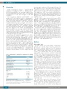
Table 1. Characteristics of 83 mantle cell lymphoma cases entering the study.
as the strongest predictor of OS in a large MCL series.20 In this context, a cohort of 20 proliferation-associated genes constructed on the basis of gene expression analysis was demonstrated to be superior to other molecular markers.20 Since approaches based on microarray technology have not yet been incorporated into routine clinical practice, a PCR-based surrogate method investigating expression of five genes has been proposed and applied to paraffin- embedded tissues.18
Recent evidence suggests that the B-cell receptor (BCR) pathway may contribute to the pathogenesis of several histological types of B-cell non-Hodgkin lymphomas, including MCL.27-30 The importance of BCR signaling path- way in B-cell malignancy pathogenesis has driven interest in the use of small-molecule inhibitors of BCR-associated kinases, potentially preventing the activation of one or more of the distal BCR signaling pathway proteins.28,31
In the present study, we developed a survival predictive model for younger patients with advanced MCL treated in the context of the Fondazione Italiana Linfomi (FIL) MCL- 0208 Phase III randomized clinical trial. This model is based upon the quantitative evaluation of six genes, most- ly from the BCR pathway, selected from a gene expression profile (GEP) of peripheral blood (PB) MCL cells and was applied to formalin-fixed paraffin-embedded (FFPE) tissue specimens. Notably, the model predicts poor response in the context of the FIL-MCL-0208 trial.
Methods
Primary MCL cases
The study included 83 out of 300 samples of adult patients under 66 years of age with advanced stage MCL, enrolled in the FIL-MCL-0208 prospective, multicenter, Phase III randomized clinical trial (clinicaltrials.gov identifier: 02354313),32 divided as fol- lows: i) a panel of 27 PB samples utilized for GEP upon positive sorting of the clonal CD5+/CD19+ MCL cells; ii) an additional panel of 19 PB samples utilized for quantitative real-time PCR (qRT-PCR) of the identified gene signature in the purified MCL cell component; iii) a panel of 43 lymph node (LN) samples uti- lized for qRT-PCR of the identified gene signature (in this LN panel 6 samples had a matched PB sample). The clinical and histo- pathological details of the 83 MCL cases used in this study are reported in Table 1. No significant differences were found between the 83 cases entering the study versus the 217 remaining cases enrolled in the clinical trial in terms of median age, MIPI score, Ki-67 index and PFS intervals (Online Supplementary Table S1 and Online Supplementary Figure S1). No differences in clinical and biological parameters were observed between PB and LN MCL samples (data not shown). All patients were treated according to the FIL-MCL-0208 clinical trial, as reported in Online Supplementary Figure S2.
Mantle cell lymphoma diagnosis was prospectively confirmed by centralized histological review according to the World Health Organization (WHO) 2008 criteria.3,33 All patients provided informed consent in accordance with Institutional Review Board requirements (0016331-BZ 09/02/2010) and the Declaration of Helsinki, and protocol consent included use of MRD sample left- overs for the study.
All the procedures employed for RNA extraction, GEP and downstream analyses, qRT-PCR, analyses and qRT-PCR valida- tions were carried out according to standard protocols, as report- ed previously.34-37 (See the Online Supplementary Appendix for details). Microarray data are available in Gene Expression
Number of cases
Median age, years (range)
Ratio male/female (%)
Abnormal LDH (%)
Median WBC (x109/L)
Typical morphology
Blastoid morphology
Median Proliferation Index (Ki-67 staining), % MIPI-c class
Low Low/intermediate High/intermediate High
na
Median survival, months (range)
Median progression-free survival, months (range)
Samples
83
56 (28-65) 57/26 (68) 45/29 (39) 13.7
74
5
20.0 (0-99)
38 (46%) 23 (28%) 11 (13%) 6 (7%)
5 (6%) 34.7 (1.4-73.4) 31.3 (1.4-73.4)
850
LDH: lactate dehydrogenase; WBC: white blood count; MIPI-c: MCL:-International Prognostic Index-combined; na: not available.
haematologica | 2018; 103(5)
