
Page 76 - Haematologica-April 2018
P. 76
A. Thivakaran et al.
A
B
C
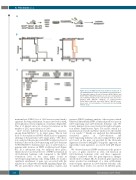
Figure 5. Loss of Gfi1b increases the stemness of LSCs. (A) In vivo limiting dilution assay for determination of functional LSCs. The indicated numbers of poly (I:C)-treated Gfi1bfl/flMxCrewt and Gfi1bfl/flMxCretg MLL-AF9 transduced cells were retransplanted into sublethally irradiated mice. (B) Survival of the mice trans- planted with different numbers of poly(I:C)-treated Gfi1bfl/flMxCrewt MLL-AF9 and Gfi1bfl/flMxCretg MLL-AF9 trans- duced cells. (C) Determination of functional LSCs by limiting dilution assay.
620
ment analysis (GSEA), loss of Gfi1b was associated with a signature showing enrichment of genes involved in AML development as well as regulation of stemness (Figure 6B). This is of interest since we observed an increase in the number of LSCs upon deletion of Gfi1b.
Gfi1b recruits different histone-modifying enzymes, among them HDACs,16 to its target genes. This in turn leads to deacetylation of H3K9, which leads to epigenetic silencing of the particular Gfi1b target genes.16 We, there- fore, analyzed the genome-wide H3K9 acetylation level of leukemic blasts from Gfi1b-expressing and Gfi1b-deficient NUP98/HOXD13tg leukemic mice. Loss of Gfi1b leads to a genome-wide increase in H3K9 acetylation level (Figure 6C). In a subsequent step, we analyzed those genes, which showed an elevated level of H3K9 acetylation in Gfi1b-deficient leukemic cells compared to the H3K9 acetylation level of the same genes found in Gfi1b-expressing leukemic cells. Using GSEA, we found a significant enrichment of gene sets associated with the regulation of cell growth and MAPK signaling (Figure 6D). We also performed a Kyoto encyclopedia of genes and
genomes (KEGG) pathway analysis of those genes, which exhibited differentially H3K9 acetylated promoter areas in Gfi1b-expressing and Gfi1b-deficient leukemic cells. We found a number of processes involved in erythroid regula- tion (Online Supplementary Figure S7A and B), which is a main function of Gfi1b and hence underscores the validity of our results.6,9,16 Finally, we analyzed the differentially acetylated genes in Gfi1b-deficient and Gfi1b-expressing leukemic cells and compared these gene sets based on the gene sets provided by the Molecular Signatures Database (MSigDB). Using this approach, we repeatedly found signatures associated with p38 (Figure 6E).
We observed increased H3K9 acetylation of the promot- er area of genes involved in stem cell function in Gfi1b-deficient leukemic cells, and these epigenetic changes correlated with the gene-expression changes described above (Figure 6B). As described, gene expression arrays revealed an enrichment of a stem cell/leukemic stem cell gene signature in Gfi1b-deficient leukemic cells. To validate these results, we selected 14 different genes
haematologica | 2018; 103(4)
