
Page 34 - Haematologica-April 2018
P. 34
578
F. Pasquier et al.
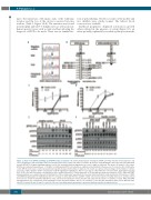
ing to the truncation of 64 amino acids of the wild-type receptor and the loss of the six last conserved tyrosine residues (Table 1, Figure 1A,B). The mutation was found in neutrophils and CD3+ T lymphocytes as well as in non- hematopoietic tissues such as nails and hair allowing the diagnosis of PFCP to be made. There was no familial his-
AB
tory of polycythemia. The blood counts of the mother and two children were strictly normal. The father’s blood count was not available.
Erythroid progenitors displayed autonomous growth when cultured in the presence of serum (Figure 1C), an effect probably explained by residual erythropoietin in the
CD
EF
Figure 1. Study of the EPOR c.1300dup (p.Gln434Profs*11) in primary cells. (A) Electropherograms showing the EPOR c.1300dup mutation in hematopoietic cells (CD3+ T-lymphocytes and neutrophils) and in non-hematopoietic tissues (nails, hair and buccal swab). (B) Scheme of EPOR wild-type and of the new frameshift EPOR mutants (EPOR FS). Negative signaling regulators (on the left) and internalization/degradation sites (on the right) are indicated. The tyrosine (Y) number of the mature EPOR is also indicated in parentheses. (C, D) Effect of erythropoietin (EPO) concentration on erythroid colony formation in the presence (C) or absence (D) of fetal bovine serum. BFU-E colonies were counted at day 14. Each experiment was performed twice in duplicate. The results are expressed in percentages of the number of colonies at 1 U/mL of EPO. (E) Effect of EPO concentration on EPOR signaling in erythroblasts. After 7 to 10 days in culture with stem cell factor (SCF), interleukin-3 (IL3) and EPO, CD34+ cells from the patient or healthy donors were cytokine-starved for 5 h then stimulated for 15 min with increasing concentrations of EPO. JAK2 and STAT5 phosphorylations were examined by western blotting. One of three independent experiments is presented and fold activation is indicated below. (F) Persistence of JAK2 and STAT5 phosphorylation in erythroblasts. After 7 to 10 days in culture with SCF, IL3 and EPO, CD34+ cells from the patient or healthy donors were cytokine-starved for 5 h prior to 15 min of stimulation with EPO 1 U/mL. Cells were then washed to remove the EPO and cultured in the absence of cytokine or serum. JAK2 and STAT5 phosphorylations were examined by western blotting in a time-dependent manner: after 5 h of starvation (-), after the EPO stimulation (+EPO) and at different times after EPO removal (10 min, 30 min, 1 h and 4 h). One out of two independent experiments is presented and fold activation is indicated below.
haematologica | 2018; 103(4)
