
Page 33 - Haematologica-April 2018
P. 33
Erythropoietin receptor mutations in PFCP
three times in phosphate-buffered saline 1X to remove the cytokine.
Signaling studies were performed using polyclonal antibodies against the phosphorylated forms of JAK2 (Tyr 1007/1008), STAT5 (Tyr 694), ERK1/2 (Thr 202/Tyr 204) and Akt (Ser 473) and against the pan proteins (Cell Signaling Technology, Ozyme, Montigny - le-Bretonneux, France). β-actin (Sigma, Saint Quentin-Fallaviers) was used as a loading control.
For glycosidase digestion, cell lysates were incubated with either endoglycosidase H (Endo H) or peptide:N-glycosidase F (PNGase F) (1,000 U) at 37°C for 16 h according to the recommen- dations of the supplier (New England Biolabs, Evry, France).
Dimerization of human erythropoietin receptor monomers assessed by split Gaussia luciferase assay.
A split Gaussia subunit, Gluc1 or Gluc2, was fused to the C-ter- minus of each EPOR construct.33 When an EPOR monomer with a Gluc1 fusion subunit dimerizes with another EPOR monomer with a Gluc2 fusion subunit, the two Gluc subunit proteins recom- bine into a catalytically active luciferase that is able to degrade coelenterazine, thus emitting light. Both EPOR constructs fused to a C-terminal Gluc1 and a Gluc2 subunit were transiently transfect- ed at a 50:50 ratio into HEK cells using Transit-LT1 (Mirus, Euromedex, Souffelweyersheim, France) as a transfecting agent. The pGL3-control vector (Promega, Charbonnières-les-Bains, France) that constitutively expresses the firefly luciferase was co- transfected in each condition as a transfection control reporter. After 48 h, luciferase signals were read by a GloMax discovery system (Promega) after addition of coelenterazine and firefly luciferin to each well. A 530LP filter was used to discriminate the luminescence of the firefly luciferase from that of the Gaussia luciferase.
Erythropoietin receptor stability assay
Ba/F3 cells were incubated for different periods of time with 50 ng/mL cycloheximide (Sigma). After cell lysis, western blotting was performed with anti-HA antibody. HA-tagged EPOR was quantified with Image J software, using β-actin as a loading con- trol. The half-life of the receptor was calculated using GraphPad PRISM software.
Cell-surface localization of HA-tagged erythropoietin receptor by flow cytometry
Cells were labeled with monoclonal mouse anti-HA antibody conjugated to phycoerythrin (Miltenyi Biotecs) and processed by flow cytometry (FACSCanto, Beckton-Dickinson, Le Pont-de- claix, France).
Erythropoietin labeling and binding and erythropoietin receptor internalization studies
Erythropoietin labeling using IODO-GEN (Pierce, Rockford, IL, USA), erythropoietin binding and EPOR internalization studies were performed as previously described.34-36 Nonspecific binding was determined using a 250-fold excess of unlabeled erythropoietin and was less than 5% in each case. All reported data represent specific binding. The number of cell-surface receptors was determined by comparing the radioactivity of Ba/F3-EPOR cells to the radioactivity of the reference UT-7 cell line.34 For internalization experiments, after incubation with 125I- erythropoietin, 5 x 10⁶ cells per condition were washed twice at 4°C to remove unbound ligand. An acidic wash was then per- formed to separate cell surface–bound from internalized ery- thropoietin. Cells were incubated in 0.5 mL acidic buffer (150 mM NaCl, 50 mM sodium acetate, pH 3.5) for 3 min at 4°C. The pH was then adjusted to 7.4 using 1 M Tris-HCl, pH 9 and the cell suspension was centrifuged. The radioactivity of the supernatant (cell surface–bound erythropoietin) and of the cell pellet (internalized erythropoietin) was determined. When 125I- erythropoietin was bound to the cells at 4°C to inhibit erythro- poietin internalization, more than 95% of cell-bound 125I-ery- thropoietin was recovered in the acidic wash supernatant using this method. Each experiment was performed three times with similar results.
Results
Identification of a new germline EPOR mutation responsible for marked erythropoietin hypersensitivity
Primary polycythemia was diagnosed in a 28-year old woman without a history of thrombosis. She had high hemoglobin concentration (21 g/dL) and hematocrit (60%) and an increased red cell mass (65%) with a low erythro- poietin level (1.2 mU/mL; laboratory standard: 5-25 mU/mL). She had no splenomegaly at physical examina- tion. Leukocyte and platelet counts in the peripheral blood as well as bone marrow aspiration and biopsy were strict- ly normal. Later no JAK2V617F or JAK2 exon12 mutation was found, rendering the diagnosis of polycythemia vera unlikely. The search for abnormal hemoglobin affinity and for VHL, PHD1/2, SH2B3 (LNK) pathological muta- tions was negative. EPOR sequencing identified a new germline heterozygous frameshift mutation, c.1300dup (p.Gln434Profs*11), which generates a new ten-amino acid C-terminal tail and a stop codon at position 444, lead-
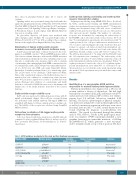
Table 1. EPOR mutations investigated in this study and their functional consequences.
Mutation (DNA)
c.1195G>T
c.1273G>T c.1311_1312del c.1327_1329delinsTAA c.1300dup
c.1330G>T
(EPOR STOP)
Mutation (protein)
p.Glu399*
p.Glu425*
p.Pro438Metfs*6
p.Pro443*
p.Gln434Profs*11 (EPOR FS)
p.Gln444*
Truncation (number of AA lost)
109
83 65 65 64
64
Number of remaining tyrosines
1
1 2 2 2
2
Reference
Arcasoy et al.11 Kralovics et al.10 Bento et al.25 This study This study
This study
EPOR: erythropoietin receptor gene (NM_000121.3), AA: amino acids.
haematologica | 2018; 103(4)
577
