
Page 54 - Haematologica3
P. 54
X. Fang et al.
miR-144/451−/− erythroblasts, AMPK is activated by over- expressed Cab39 and promotes apoptosis by inhibiting mTOR activity. It is unclear how levels of stress affect the regulatory role of AMPK in apoptosis, i.e. does AMPK favor cell survival under mild stress but enhance apoptosis under severe stress?
In this study, we found that loss of miR-144/451 induces apoptosis in a p53-dependent fashion. Some evidence from a study in Diamond-Blackfan anemia (DBA) has shown that haploinsufficiency of ribosomal protein sub- units can induce erythroblast apoptosis via p53-dependent mechanisms.37 Stabilization of p53 by the MDM2 antago- nist SAR405838 induces major hematopoietic defects including erythroid precursor apoptosis in vitro.38 How p53 is activated after miR-144/451 depletion remains to be elu- cidated. In some cell types, AMPK induces apoptosis by activating p53 through phosphorylation,34,35 whereas oth- ers have reported that p53 activates AMPK/mTOR signal- ing to suppress cell growth by targeting sestrin 1 and ses- trin 2 upon stress.39 In addition, several groups report an inhibitory role of mTOR activity on p53 function.28, 29 It is possible that a positive feedback loop involving AMPK, p53, and mTOR signaling regulates apoptosis of miR- 144/451−/− erythroid cells under stress conditions, although further investigations are required to fully define the process.
Anemia is a common complication after orthotopic kid- ney transplantation due to multifactorial effects including iron deficiency, reduced erythropoietin production, and chronic or acute inflammation. Additionally, mTOR inhibi-
tion by sirolimus is a possible risk factor for the develop- ment of anemia in kidney transplant recipients.40,41 There is also a substantial risk of anemia from the mTOR inhibitor everolimus in cancer therapy.42 Interestingly, studies in ani- mal models and cell cultures demonstrate that treatment with mTOR inhibitors reduces RBC size, independent of alterations in kidney function.32,43 Consistent with this find- ing, miR-144/451−/− mice exhibit microcytic anemia,15 per- haps due to mTOR inhibition. Moreover, the current study shows that these mice exhibit enhanced erythroblast apop- tosis with various erythropoietic stresses including devel- opmental expansion of FL, hemolysis, acute blood loss, and precursor depletion by 5-FU, a chemotherapeutic drug. Accelerated apoptosis in the absence of miR-144/451 is caused by overexpression of the miR-451 target Cab39, which activates AMPK, thus inhibiting mTOR signaling. Therefore, our data explain why erythroid apoptosis is an underlying mechanism of profound anemia when the enzymatic activity of Cab39/LKB1/AMPK is significantly increased and mTOR signaling is perturbed.
A mechanistic understanding of the differences between steady state and stress erythropoiesis could be of benefit in multiple clinical settings.44 Moreover, these processes are likely to be impacted differently by various disease states. Common causes of acquired anemia include dietary iron deficiency, malaria, chronic infectious dis- eases, autoimmune or rheumatological disorders, chemotherapy, and chronic kidney disease. Genetic caus- es of anemia include DBA and hemoglobinopathies such as sickle cell disease (SCD) and thalassemia.45 Of note,
A
C
B
D
414
E
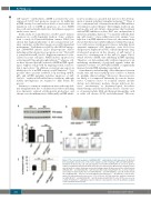
Figure 7. The increased apoptosis of miR-144/451−/− erythroblasts is p53-dependent. (A) Western blot showing the protein levels of the tumor suppressor p53 in wild-type (WT) and miR-144/451−/− (KO) E14.5 fetal liver (FL) cells. Lineage negative-selected WT and KO E14.5 FL cells were grown in culture in erythroid maturation medium, and cells were harvested for Western blot analysis for p53 after 24 hours in culture. Actin was used as the loading control. (B) Gross view of the cell pellets from miR-144/451−/− and miR-144/451−/−/p53 knock-in (KI) double-mutant E14.5 FLs. (C) Total erythrob- last number for whole E14.5 FLs from miR-144/451−/− and miR-144/451−/−/p53 KI double-mutant mice. miR-144/451−/− mice were crossed with p53-deficient KI mice.16,31 N=3 WT mice, n=6 KO mice, and n=6 double-mutant mice were used. **P<0.01 (t-test). (D) Flow cytometric analysis of apoptosis for miR-144/451−/− and miR-144/451−/−/p53 KI double-mutant E14.5 FL cells. WT FLs were used as controls. PI−Annexin V+ labeling was taken to indicate early apoptotic cells. (E) Quantitative analysis of flow cytometric data from (D).
haematologica | 2018; 103(3)
