
Page 37 - Haematologica3
P. 37
Modeling human erythroid-macrophage interactions
Cytospins
Cells were cytospun using Shandon Cytospin II (Thermo Scientific), dried and fixed in methanol. Cells were stained with benzidine and Differential Quik Stain Kit (PolySciences, Warrington, PA, USA) following manufacturer’s instructions. Slides were dried, embedded in Entellan (Merck, Darmstadt, Germany) and images were taken (Leica DM-2500, Germany).
Reverse transcription polymerase chain reaction analysis
Reverse transcription polymerase chain reaction (RT-PCR) was performed as previously described.12 Values were normalized using S18 and HPRT as a reference gene and calibrated relative to expression of CD14+ monocytes at day 0 (primers listed in Online Supplementary Methods).
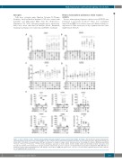
AB
CD
E
Figure 1. Glucocorticoid receptor activation directs CD14+ monocytes towards a tissue resident macrophage phenotype. (A-D) Distribution graphs displaying the relative geometric mean fluorescence intensity (MFI) of CD16, CD163, CD169 and CXCR4 on human monocytes (n=3-6) cultured for three days under various con- ditions (EPO, SCF, lipids or dexamethasone). MFI was normalized to change to isotype control and presented as fold change (fc). Mean ± SEM (two-way ANOVA, *P<0.05, **P<0.01, ***P<0.001, ****P<0.0001). (E) Relative expression of CD16, CD163, CD169, CXCR4, CD206 and DC-SIGN on CD14+ monocytes (n=3) directly after isolation from mononuclear cells (D0) and after culture in the presence or absence of dexamethasone (Dex) and/or mifepristone (Mif). MFI was nor- malized to isotype control and displayed as a fold change to day 0. Mean ± SEM (ratio paired t-test, *P<0.05, **P<0.01). EPO: erythropoietin; SCF: stem cell factor; ns: not significant; ND: not detected.
haematologica | 2018; 103(3)
397
