
Page 75 - 2020_08-Haematologica-web
P. 75
Exposure to 20 mGy radiation decreases HSC functions
Low doses of ionizing radiations do not induce DNA double strand breaks nor activate ATM or p53 signaling pathway
Since irradiation usually induces DNA double strand breaks (DSB), we quantified the number of H2AX and 53BP1 foci 30 min post irradiation (Figure 4A). In contrast to a 2.5 Gy irradiation, a 20 mGy irradiation did not increase the number of H2AX and 53BP1 foci compared to sham- irradiated CD34+CD38lowCD45RA–CD90+ HSPC indicating that 20 mGy LDIR does not induce DNA DSB (Figure 4B). We then studied the DNA damage response (DDR) path-
way after exposure to LDIR by quantification of ATM and p53 phosphorylation 10 min and 3 h after irradiation. As expected, 2.5 Gy-irradiated HSPC exhibited an increased ATM and p53 phosphorylation compared to control HSPC (Figure 4C and D). In contrast, no increase in ATM or p53 phosphorylation was detected after exposure to 20 mGy (Figure 4C and D). Importantly, the expression of p53 pro- tein was not modified by IR (Online Supplementary Figure S3). Altogether these results indicate that 20 mGy LDIR does not induce DNA DSB nor activate p53 and ATM-dependent DNA damage repair pathway in human HSPC.
AB
CD
E
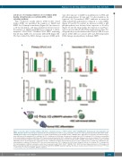
Figure 6. Low doses (LD) of ionizing radiations (IR) induce a transitory increase of ROS in CD34+ CD38low CD45RA–CD90+ hematopoietic stem progenitor cells (HSPC) that alters their serial clonogenic potential. (A) Colony forming unit-cell (CFU-C) assay. Cumulative results from 3 independent experiments with CD34+ CD38low CD45RA–CD90+ HSPC from 3 independent pools of cord blood (CB) samples. Sorted CD34+ CD38low CD45RA– CD90+ HSPC were pre-treated or not with N-acetylcys- teine (NAC) prior to IR and plated (500 cells/plate) in CFU-C conditions for 12-14 days. Shown are the number (nb) of CFU-C (primary CFU-C). Results are normalized to the sham-irradiated conditions. (B) Primary CFU-C were pooled and replated in CFU-C conditions for 12-14 days. Shown are the nb of secondary CFU-C, normalized to the sham-irradiated conditions (cumulative results from 3 independent experiments). (C) Sorted CD34+ CD38low CD45RA– CD90+ HSPC were pre-treated or not with SB203580 prior to IR and plated (500 cells/plate) in CFU-C conditions for 12-14 days. Shown are the nb of CFU-C (primary CFU-C). Results are normalized to the sham-irradiated conditions. (D) Primary CFU-C were pooled and replated in CFU-C conditions for 12-14 days. Shown are the nb of secondary CFU-C, normalized to the sham-irradiated conditions (cumulative results from 2 experiments with CD34+ CD38low CD45RA– CD90+ HSPC from two independent pools of CB samples. (E) Model explaining how LDIR can impair HSC self-renewal through ROS-p38MAPK dependent pathway. Results are shown as mean+standard error of mean. **P<0.01, ***P<0.001, ****P<0.0001 (Mann- Whitney statistics).
haematologica | 2020; 105(8)
2051
