
Page 72 - 2020_08-Haematologica-web
P. 72
E. Henry et al.
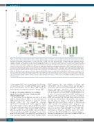
A
B
CD
Gated in Linneg CD34+ CD90+ subset
EF
Figure 3. Low doses (LD) of ionizing radiations (IR) do not induce apoptosis and do not modify the cell cycle in human hematopoietic stem progenitor cells (HSPC).
(A) CD34+ cells were irradiated and cultured for 6 hours (h) at 37°C, then stained for cell surface markers and fixed. Cleaved-caspase 3 protein expression was ana- lyzed by FACS. Percentage of cleaved-caspase 3+ cells on CD34+ CD38low CD45RA– CD90+ HSPC and on total CD34+ (left panel) and overlay histograms of cleaved- caspase 3 expression on HSPC (right panel) are represented in function of IR doses. One representative experiment out of two is shown. (B) Sorted CD34+ CD38low CD45RA–CD90+ HSPC were irradiated or not and co-cultured with MS5 stroma cell line for several days. At several time points, cells were numerated and stained for cell surface markers. The numbers of CD34+ cells (left) and LinnegCD34+CD90+ cells (right) were monitored over time. One representative experiment out of two is shown. (C-F) Sorted CD34+ CD38low CD45RA– CD90+ HSPC were first stained with carboxyfluorescein hydroxysuccinimidyl ester (CFSE), irradiated and cultured for sev- eral days. One representative experiment out of two is shown. (C) Differentiation of CD34+ CD38low CD45RA– CD90+ HSPC in culture was followed by using expression levels of CD90 and CD34 surface markers. Dot plots (left panel) represent CD90 and CD34 expression after 2 and 8 days of culture for control and 20 mGy-irradiated HSPC. Histogram bars (right panel) represent the percentage of LinnegCD34+CD90+ cells at different days of culture after IR. (D) Levels of carboxyfluorescein succin- imidyl ester (CFSE) fluorescence in the LinnegCD34+CD90+ subset at different days of culture in control and 20 mGy conditions. No differences in cell division can be detected between both conditions. (E) Histogram representing CFSE staining in the HSPC-derived bulk cells at days 6 and 8 of culture in control and 20 mGy condi- tions (left panels). Histogram bars show CFSE labeling loss over culture time in the bulk population (right panel). (F) Percentage of LinnegCD34+CD90+ cells in CFSEhi cells in control and in 20 mGy conditions. Results are shown as mean±standard error of mean. **P<0.01, ***P<0.001 (Mann and Whitney statistics). Abs Nb: absolute numbers.
of the engrafted HSC was shown (Figure 2D) showing a cell-autonomous effect of LDIR in HSPC. Taken together these results indicate that 20 mGy LDIR affects the hematopoietic reconstitution capacity of human HSPC.
Low doses of ionizing radiations do not induce apoptosis nor alter cell cycle of hematopoietic stem/progenitor cells
To characterize the mechanisms activated by 20 mGy LDIR in human primary HSPC, we first investigated if LDIR trigger apoptosis. Analysis of caspase 3 cleavage showed that 2.5 Gy irradiation increases CD34+CD38lowCD45RA–CD90+ HSPC apoptosis (Figure 3A, red histograms), whereas 20 mGy LDIR had no signif- icant effect on the percentage of cleaved caspase 3-express- ing HSPC compared to sham-irradiated HSPC (Figure 3A, green histograms). As increased HSPC cell cycle can lead to self-renewal defects and HSC exhaustion,28 we also deter- mined if 20 mGy LDIR could alter HSPC proliferation. 20 mGy and sham-irradiated CD34+CD38lowCD45RA–CD90+
HSPC generated the same number of CD34+ and CD34+CD90+ cells in vitro (Figure 3B, left and right panel, respectively) whereas 2.5 Gy-irradiated HSPC did not pro- liferate efficiently. Moreover, the proportion of CD34+CD90+ cells evolved similarly during culture period after no irradiation or 20 mGy LDIR, strengthening the lack of effect of a 20 mGy irradiation on HSPC differentiation (Figure 3C). We studied the cell division rate of irradiated HSPC after staining with CFSE and during culture in serum free medium with cytokines. The number of cell divisions was the same for sham- or 20 mGy-irradiated HSPC (Figure 3D). After 6 and 8 days of culture, no difference in CFSEhi cell (the most immature cells)28 proportion was observed between 20 mGy and control HSPC (Figure 3E) and 90% of these CFSEhi cells expressed the immature markers CD34 and CD9029 regardless of the culture time and irradiation conditions considered (Figure 3F). These results show that 20 mGy LDRI does not impair either pro- liferation or differentiation of the most immature HSPC in cytokine-stimulating conditions.
2048
haematologica | 2020; 105(8)
