
Page 31 - 2020_08-Haematologica-web
P. 31
Inherited thrombocytopenias
the degree of thrombocytopenia because the absence of GPIba prevents the shear-dependent tethering of platelets to von Willebrand factor (VWF) in vascular lesions, an abnormality mimicked by the loss of ristocetin-induced platelet agglutination in a diagnostic test.20,21 The search for mutations first targeted GP1BA, the gene encoding GPIba, but mutations in GP1BB and GP9 were also quickly shown to cause BSS by preventing the surface expression of GPIba.22 The generation and rescue of BSS in a mouse model confirmed the link between GPIba loss and the appearance of giant platelets and, therefore, macrothrom- bocytopenia.23 Typical findings in BSS and the mouse models are aberrant formation of the demarcation mem- brane system in megakaryocytes while fewer proplatelets protrude into the vascular sinus and these proplatelets are thicker with larger heads. While the loss of megakaryo- cyte interactions with extracellular proteins remains a plausible molecular basis of BSS, the absence of mechani- cal stabilizing interactions between GPIb, cytoskeletal proteins and internal membranes is another likely factor.
While classic BSS has autosomal recessive (AR) inheri- tance, mono-allelic forms with autosomal dominant (AD) transmission are a frequent cause of mild macrothrombo- cytopenia in Europe. The initial example was the Bolzano (p.A172V) mutation affecting GPIba in Italian families, said to be responsible for Mediterranean macrothrombo- cytopenia, although other variants of GP1BA have since been described. More recently a series of single allele vari- ants of GPIBB have been identified by whole exome sequencing (WES) in patients with mild macrothrombocy- topenia.24 The difference in phenotype given by AD single
allelic forms of BSS compared with heterozygosity for bi- allelic BSS has yet to be fully explained. A 1.5 to 3.0-Mb hemizygous mostly somatic deletion on chromosome 22q11.2 including GPIBB is seen in the Di-George and velocardiofacial syndromes in which multiple develop- mental defects are often accompanied by mild to moder- ate macrothrombocytopenia.21
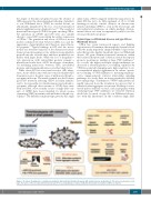
Figure 3. A cartoon showing genes causing non-syndromic and syndromic thrombocytopenias with normal sized or small platelets. The genes are grouped accord- ing to the nature of the encoded abnormal protein and to the accompanying secondary condition(s) that define the syndrome. BM: bone marrow.
Platelet-type von Willebrand disease and type 2B von Willebrand disease
GPIba has seven leucine-rich repeats and flanking regions near its N-terminus; the mucin-like domain follows with the many negatively charged O-linked oligosaccha- rides that provide rigidity. In platelet-type von Willebrand disease, AD gain-of-function missense mutations within the leucine-rich repeats (and 1 deletion outside the repeats) promote spontaneous binding of large VWF multimers.25 As a result, the higher molecular weight multimers are decreased or absent in plasma. Cross-linking of platelets by VWF favors platelet clumping and a high sensitivity to ris- tocetin-induced platelet agglutination. In culture, sponta- neous binding of VWF multimers to maturing megakary- ocytes inappropriately activates intracellular signaling pathways; as a result, there are fewer proplatelets and they have enlarged tips.26 Furthermore, VWF-bound platelets are rapidly removed from the circulation in a process that is enhanced when aggregates are present. Bleeding is accen- tuated under conditions of stress, such as pregnancy when circulating large VWF multimers are elevated. Mention should also be made of type 2B von Willebrand disease, also with AD inheritance. In this condition single allele
haematologica | 2020; 105(8)
2007
