
Page 29 - 2020_08-Haematologica-web
P. 29
Inherited thrombocytopenias
some glycolipids.5 The discovery that a major component of the platelet surface, first identified as “GPI”, was absent from platelets in BSS began the long road to defining the molecular landscape of inherited thrombocytopenias.6 This centenary review will take a concise look at this jour- ney emphasizing major steps in the current understanding of these conditions and highlighting recent advances in what is an intriguing subset of Mendelian diseases.
Platelet production and lifespan
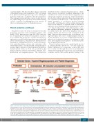
The platelet count is the result of a balance between the biogenesis, senescence and consumption of these cells. Briefly, CD34+ hematopoietic stem cells (HSC) first give rise to megakaryocyte-erythrocyte progenitors, a propor- tion of which will develop into colonies that mature by endomitosis to polylobulated megakaryocytes with a high DNA content (Figure 1). Megakaryocyte differentiation is regulated by transcription factors as well as by interac- tions with chemokines, cytokines and constituents of the extracellular matrix.7 Thrombopoietin, acting through its receptor Mpl, is a key factor.8 Mostly produced in the liver, thrombopoietin levels in blood are governed by the platelet number; feedback mechanisms stimulate HSC proliferation and megakaryopoiesis. The demarcation
membrane system of mature megakaryocytes is a mem- brane reservoir for platelet biogenesis. Migrating to the endothelial cell sinusoid barrier, mature megakaryocytes extend long, branched, protrusions, termed proplatelets, into the blood where under flow they release large num- bers of platelets from their ends.9,10 This whole process is highly dependent on cytoskeletal proteins including myosin IIA, actin filaments and the tubulins that provide tracks for the transport of organelles with a Cdc42/RhoA regulatory circuit guiding transendothelial platelet biogen- esis.11 Type I collagen in the extracellular matrix is a nega- tive regulator, preventing premature proplatelet forma- tion. Early reports also insisted that megakaryocytes can migrate directly into the blood and accumulate in the lungs. Using two-photon microscopy, in vivo lineage trac- ing technologies and a series of lung transplants in mouse models, Lefrancais et al.12 have confirmed that the lung is indeed a site for platelet biogenesis. In conditions of acute need, direct megakaryocyte rupture is an alternative path- way for platelet production.13
If unused in hemostatic events or pathological process- es, human platelets circulate for 7-10 days (Figure 2). Platelet clearance due to senescence occurs in the liver. Loss of sialic acid through the action of neuraminidase during platelet aging exposes galactose or N-acetyl-galac- tosamine, residues that mediate platelet binding to the
Figure 1. A schema of the major steps of megakaryopoiesis highlighting how inherited defects of selected genes cause thrombocytopenia. Megakaryocytes arise from hematopoietic stem cells that proliferate to first form megakaryocyte-erythrocyte progenitors, a proportion of which give rise to colony-forming megakaryocyte progenitors. These events require the interaction of thrombopoietin with its receptor and are under the influence of various growth factors (e.g. stem cell factor, cytokines and interleukins). The developing megakaryocytes undergo endomitosis to increase chromosome number; they then mature before migrating to the vas- cular sinus where they extend proplatelets or migrate themselves across the endothelial cell barrier into the vascular sinus. This latter step is under the influence of many factors including stromal derived growth factor-1 and sphingosine-1-phosphate. Selected genes with variants causing inherited thrombocytopenias are shown in red. HSC: hematopoietic stem cells; TPO: thrombopoietin; MPL: thrombopoietin receptor; MEP: megakaryocyte-erythrocyte progenitor; SCF: stem cell factor; IL: interleukin; MKP: megakaryocyte progenitor, MK: megakaryocyte; SDF-1: stromal derived growth factor-1; S1P: sphingosine-1-phosphate; PPL: proplatelet.
haematologica | 2020; 105(8)
2005
