
Page 204 - 2020_08-Haematologica-web
P. 204
D. Stefanoni et al.
Storage-induced lipid remodeling and oxidant damage of membrane lipids is higher in Rhesus macaque, as compared to human, red blood cells
Interestingly, untargeted analyses revealed significantly higher levels of diethylhexyl-, monoethylhexyl- and free phthalate plasticizers in RM RBC as a function of storage (Online Supplementary Figure S3). Since both human and RM RBC samples were stored in the same polyvinylchlo- ride units under identical conditions, we hypothesize that these results could, at least in part, be explained by differ- ential species-specific storage-dependent membrane dynamics. Further analyses of targeted metabolomic data indicated that RBC acylcarnitines increased with storage duration. RM RBC were characterized by higher levels of short and medium chain acylcarnitines (C2-12), and lower levels of long and very-long acylcarnitines (C16-22
or longer), as compared to human RBC (Figure 6). Similarly, free fatty acids (medium and long-chain, but not very long-chain fatty acids) were higher in RM RBC (Figure 6), along with higher levels of sphingosine 1-phos- phate and lipid peroxidation products, including 4- hydroxynonenal (4-HNE) and its glutathionylated form (GS-HNE). Consistent with increased oxidant stress in stored RM RBC, along with tracing experiments in fresh RBC (Figure 2), RM RBC showed significantly higher storage-dependent increases in lactoyl-glutathione levels (Figure 6).
To expand on these observations, untargeted lipidomic analyses were performed on fresh (day 0) and end of stor- age (day 42) RM and human RBC (summarized by lipid classes in Online Supplementary Figure S5 and volcano plots in Online Supplementary Figure S6A, B). Notably,
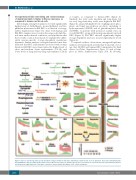
Figure 4. Species- and storage time-specific metabolic changes in Rhesus macaque and human red blood cells: focus on glycolysis, the pentose-phosphate path- way, glutathione, and one-carbon homeostasis. Data for Rhesus macaques are shown in green, those for humans are represented in red. Supernatant metabolites are shown in the right-hand side of the figure, outside the representative lipid bilayer of the cellular membrane.
2180
haematologica | 2020; 105(8)
