
Page 194 - 2020_08-Haematologica-web
P. 194
Z. Hao et al.
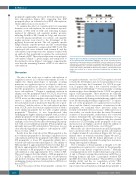
Figure 5. Effect of naturally occurring propeptide mutations at positions -1 and -4 on reporter-protein carboxylation. Wild-type and -1 and -4 mutant reporter- proteins were transiently expressed in HEK293 cells; the transfected cells were cultured with a serum-free medium containing 11 mM vitamin K (Vit. K) or 5 mM warfarin (Warfarin). The cell culture medium was collected 48 hours post-trans- fection and loaded to SDS-PAGE for Western blot analysis. Carboxylated protein bands were probed by a mouse monoclonal antibody that recognizes Gla residues.
propeptide significantly decreased (10-fold) reporter-pro- tein carboxylation (Figure 6D), suggesting that BGP propeptide plays an essential role in BGP carboxylation, which differs from in vitro studies.14,15
To examine the effect of coagulation factor’s remaining domains on its carboxylation, we used chimeric reporter- proteins of FIX with its EGF and following domains replaced by different cell organelle marker proteins, including Sec61B for ER, Giantin for Golgi, and tissue fac- tor for the plasma membrane. As controls, cell organelle marker proteins were fused to the N-terminal of the mCherry fluorescent protein. The cell organelle specific FIXgla chimeric reporter-proteins and the corresponding controls were transiently co-expressed in COS-7 cells. The transfected cells were cultured with vitamin K, and the carboxylated reporter-protein was immuno-stained with an antibody that specifically recognizes the carboxylated Gla domain of FIX. These reporter-proteins were properly carboxylated (Figure 7, green image) and transported to the destined locations (Figure 7, red image), supporting the view that the remaining domains of FIX do not affect its carboxylation.
Discussion
The aim of this study was to explore carboxylation of coagulation factors in a cellular environment in order to explain the clinical phenotypes of naturally occurring mutations in coagulation factors, as related to their car- boxylation modifications. Previous studies have shown that the propeptide is essential for directing coagulation factor carboxylation.7,13 Despite a significant variation in affinity, once the propeptide binds to GGCX, it has been proposed that it induces a conformational change in the GGCX active site that stimulates carboxylation of its sub- strate to a similar extent.23 The in vitro study shows that the carboxylation rate is much faster than the rate of prod- uct release,37 and the release of the carboxylated product from GGCX can be detected in coagulation factors with a lower affinity propeptide but not with a higher affinity propeptide.38 Therefore, it was hypothesized that exchanging the higher affinity propeptide with a reduced affinity propeptide would enhance coagulation factor car- boxylation by allowing for a higher substrate turnover. For example, it has been shown that substituting FX propep- tide with a lower affinity propeptide (PT propeptide) sig- nificantly increased FX carboxylation.17 However, it is not clear why this hypothesis applies to carboxylation of FX17 but not to that of FIX.18
Results from this study show that FIX propeptide is the most efficient propeptide for directing coagulation factor carboxylation and that the propeptide with either a higher (FX propeptide) or lower (PC propeptide) affinity has a reduced carboxylation efficiency (Figure 1). The affinity of FIX propeptide is approximately 8-fold lower than that of FX and is approximately 4-fold higher than that of PC.16,23 The efficiency of FX propeptide to direct reporter protein carboxylation is only approximately 10% of that of FIX. This result suggests that the affinity of FIX propeptide for GGCX is optimal, as it balances the rate of carboxylation and product releasing. This explains why the propeptide exchanging strategy increased carboxylation of FX but not that of FIX.17,18
It has been proposed that the propeptide contains two
recognition elements: one for GGCX recognition (located towards the N-terminus) and one for propeptidase recog- nition (located near the C-terminus). For GGCX recogni- tion, it appears that only a few conserved residues are essential for GGCX binding.25,39 Several naturally occurring mutations have been identified in the GGCX recognition region of FIX propeptide. These mutations are clinically silent in normal conditions, but selectively decrease FIX activity dramatically during warfarin therapy, which could cause life-threatening bleeding complications.27 To explore the role of the propeptide on coagulation factor carboxy- lation and the clinical consequence of mutations in the propeptide, we examined these questions using our recently established cell-based assays.40 Unlike previous in vitro studies, our results show that the entire N-terminal sequence of the propeptide, rather than a few conserved residues, determines the carboxylation efficiency of coag- ulation factors (Figure 3). This explains why the essential residues for GGCX binding in the propeptide of all coag- ulation factors are highly conserved, while the affinity of the propeptides for GGCX varies over 100-fold.16
Our results also show that mutations in FIX propeptide have a moderate effect on reporter-protein carboxylation at a higher vitamin K concentration (Figure 4A), but a sig- nificant effect on warfarin sensitivity (Figure 4B and C), which is consistent with the clinical phenotype of war- farin hypersensitivity in patients bearing these mutations during anticoagulation therapy.
The C-terminus of the propeptide is thought to be the propeptidase recognition site, essential for the propeptide cleavage to form mature coagulation factors.13 Naturally occurring mutations were found in this region at positions -4 and -1. Patients carrying these mutations in FIX have a bleeding diathesis and have a propeptide attached FIX (proFIX) detected in their plasma.30 This proFIX loses lipid binding ability and has no coagulation activity.41 Our results show that mutations at -4 and -1 do not affect reporter-protein carboxylation, although the propeptide is
2170
haematologica | 2020; 105(8)
