
Page 61 - 2020_07-Haematologica-web
P. 61
protein to form multiprotein transcription complexes that regulate the differentiation of various cell types. Ldb1 KO (Ldb1-/-) mice die between E9.5 and E10 due to severe defects in a number of developing tissues, including abnormal hematopoietic development.10 This abnormal hematopoiesis is also observed in knockout mouse embryos lacking the LDB1 binding-partners TAL111 or LMO2.12
Despite the knowledge on GATA1 binding partners, it is not known when and where GATA1 complexes form. In order to identify the temporal and spatial appearance of GATA1/FOG1 and GATA1/LDB1 complexes during differ- entiation, we applied proximity ligation assays (PLA)13 in differentiated mouse embryonic stem (ES) cells and FL cells. We detect the first significant GATA1/LDB1 interac- tion in CD71+ FL cells. Knockdown (KD) of LDB1 in vitro led to fetal cell death and decreased the CD71+ cell popu- lations, providing functional evidence for its essential role at that stage of erythroid differentiation in normal FL.
Methods
Cell culture and mouse FL collection
Wild-type (WT) and Ldb1-/- mouse ES cells were cultured in DMEM-15% FCS-1% non-essential amino acids-100 units/mL penicillin-100 mg/mL streptomycin-6.3e-4% 2-mercaptoethanol- 100 units/mL Esgro. Day 12.5 (D12.5) or D13.5 FL were used for cell sorting, nuclear extraction, or directly embedded in OCT Tissue-Tek (Sakura) for tissue slicing. All animal experiments were performed according to guidelines and protocols that had been approved by an independent committee on the ethical use of experimental animals (DEC).
ES cell differentiation by the hanging drop method
Mouse WT and Ldb1-/- ES cells were differentiated as described.14 On D4, D5 or D9 of ES cell differentiation, embryoid bodies (EB) were collected by flushing with PBS in 50 mL falcon tubes then embedded in the OCT Tissue-Tek.
Flow cytometry analysis and cell sorting
Mouse E12.5 or E13.5 FL cells (infected or not by LDB1 or GATA1 small hairpin RNA [shRNA]) were labeled with CD71-FITC and TER119-PE antibodies and sorted on a FACSAria III (BD Biosciences) into four populations: P1 (CD71–/TER119–), P2 (CD71+/TER119–), P3 (CD71+/TER119+) and P4 (CD71–/TER119+).
Real-time quantitative PCR (RT-qPCR)
Total RNA was isolated from sorted FL cells or trypsin-dissoci- ated EB up to D6 of differentiation with Trizol (Invitrogen). RT- qPCR was performed using SybrGreen (Applied Biosystem) on Bio-Rad CFX96. Rnh1 (ribonuclease inhibitor 1) gene was used as internal control for normalization. Primers are indicated in the Online Supplementary Table S1.
Gene expression profiling by RNA sequencing (RNA-seq)
RNA samples from sorted mouse E12.5 FL cells, P1 to P4, were sequenced and analyzed as described10 using independent biolog- ical replicates. Significant (at least ±0.6 log two-fold change and P-value ≤0.05) up- and down-regulated genes were selected. Data are deposited in the Sequence Read Archive (SRA) (Accession Number: SRP158286).
Antibodies
Antibodies are indicated in the Online Supplementary Table S2.
RNA interference
Lentiviral particles for LDB1 were produced as described by Stadhouders R15 using Ldb1 shRNA (shRNA#1: 5’-GGACCAAA- GAGATATACCA-3’, shRNA#2: 5’-GACTCTGTGTGATACTA- GA-3’) and Gata1 shRNA (5’-GTTTGGATGCAGCATCTTCTT- 3’) with non-targeting shRNA as controls. Lentiviral infected cells were harvested 72 hours after transduction and processed for nuclear extraction.
Protein analysis
Murine erythroleukemia (MEL) cells or EB nuclear extract and immunoprecipitation (IP) were prepared as described16 and size- exclusion chromatography was performed on an AKTA-FPLC apparatus with a Superose-6 10/30 column (Amersham Biosciences). Fractions were precipitated with trichloroacetic acid and analyzed by Western blotting using Odyssey system (LI- COR).
Immunofluorescent staining
MEL or FL cells were stained as described8 and analyzed by con- focal microscopy (Leica SP5).
haematologica | 2020; 105(7)
Temporal and spatial emergence of GATA1 complex
PLA on EB and mouse embryo tissue
10 μm sliced E4, 5, 9 EB or E12.5 mouse fetal tissues were fixed and processed for PLA following the manufacturer’s protocol (Duolink, OLINK) using antibodies indicated in the Online Supplemntary Table S2. PLA signals were visualized by Leica SP5 confocal microscopy and were analyzed using BlobFinder soft- ware (Uppsala University, Sweden). Signals contained in or adja- cent to nuclei were compared between different groups (n=3). The Kruskal–Wallis test for variance between groups was performed and the Tukey method to counteract multiple comparison errors was applied. Deconvolution of PLA signals and volume analysis was performed using Huygens Suite as published.17
Results
LDB1 complexes start to form at D4 of in vitro ES cell differentiation
We applied PLA18 on sliced in vitro differentiated EB to identify when GATA1 complexes form. This enables low level detection of endogenous protein-protein interaction in situ.
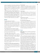
First, we characterized gene expression dynamic for genes of interest during ES cell differentiation (Figure 1A). As expected, the stem cell marker Rex1 is expressed early (day 0 to 2 [D0-D2]) and decreases during differentiation, while β-globin increases at later stages at D5-D6. Thus Ldb1 is expressed both in early and late stages of ES differ- entiation, and in the erythroid cell lineage at D5-D6. Following differentiation Gata1, Fog1, Gata2, Flk1, Tal1 and Lmo2 expression gradually increases. Of note Gata2 gene induction starts at D4 whereas Gata1 expression is delayed for 24 hours (h) (Kolovos et al. submitted), i.e. the GATA-switch occurs in early embryogenesis.19
PLA representing combinations of transcription factor (TF) interactions (GATA1/LDB1, GATA1/FOG1 and LDB1/E2A) was performed in undifferentiated cells D0, D4, D5 and D9 differentiated WT or Ldb1-KO EB (Figure 1B). Quantification of PLA signals showed that these inter- actions are absent in ESC, while GATA1/LDB1 and LDB1/E2A interactions already occur at D4 of ES cell dif- ferentiation. The GATA1/FOG1 interaction appeared 24 h later at D5. No red blood cells emerged in Ldb1-KO EB at
1803
