
Page 130 - 2020_07-Haematologica-web
P. 130
I. Truxova et al.
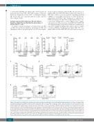
to efficiently kill K562 cells (Figure 2D), CRTHi patients in remission possessed NK cells with superior cytotoxic functions compared to their CRTLo counterparts (Figure 2E). These data are consistent with the results reported above (Figure 2A-B).
Surface-exposed CRT influences NK-cell effector functions indirectly, by affecting the phenotype of CD11c+CD14high cells
To further evaluate the impact of surface-exposed CRT on NK cells and the mechanisms underlying its NK cell- stimulatory effects, we performed a set of in vitro experi-
AB
ments with recombinant CRT (rCRT). Pre-incubation of purified NK cells with rCRT did not affect the capacity of NK cells to release cytotoxic granules containing perforin 1 (PRF1) or secrete IFN-g in response to either nonspecific stimulation with PMA and ionomycin or exposure to K562 cells (Figure 3A and Online Supplementary Figure 2A). Conversely, adding rCRT to whole PBMCs led to signifi- cant increase in the percentage of CD45+CD3–CD56+ NK cells degranulating in response to PMA plus ionomycin or exposure to K562 cells (Figure 3B), with no effects on IFN- g secretion (Online Supplementary Figure S2B). We con- firmed these results with NK-cell cytotoxicity assays, as
P=0.02 P=0.04 P=0.01 P=0.0001
CD
E
P=0.004
Figure 2. The impact of ecto-CRT on the activation and cytotoxic potential of natural killer cells in acute myeloid leukemia patients. (A, B) The percentage of IFN-γ- and degranulating (CD107a+/GZMB+) CD45+CD3–CD56+ natural killer (NK) cells upon PMA + Ionomycin or K562 cell line stimulation in 17 CRTLo and 18 CRTHi acute myeloid leukemia (AML) patients prior to the induction chemotherapy (A) or in 12 CRTLo and 12 CRTHi AML patiens after the restoration of normal hematopoiesis (B). Patient samples were analyzed by flow cytometry. Box plots: lower quartile, median, upper quartile; whiskers, minimum, maximum; ns: not significant. (C) Cytotoxic potential of NK cells isolated from AML patients before the initiation of chemotherapy (Prior, n=10) versus upon the restoration of normal hematopoiesis (Recovery, n=10). Purified NK cells were tested for their ability to kill target K562 cell line at two different effector:target cell ratios (5:1 and 10:1) and the viability of K562 cells was determined by flow cytometry after 4 hours (h). (D, E) Cytotoxic potential of NK cells isolated from five CRTHi and 5 CRTLo AML patients before the initiation of chemotherapy (D) or upon the restoration of normal hematopoiesis (E). Purified NK cells were tested for their ability to kill target K562 cell line at effector:target cell ratio 5:1 and the percentage of dead (AnnV+DAPI+) K562 cells was determined by flow cytometry after 4 h. The representative dot plots of NK cell cytotoxicity assay showing the viability of target K562 cells in CRTHi versus CRTLo AML patients before the initiation of chemotherapy (D) or upon the restoration of normal hematopoiesis (E) are shown. Box plots: lower quartile, median, upper quartile; whiskers, minimum, maximum; ns: not significant. CRT: calreticulin.
1872
haematologica | 2020; 105(7)
