
Page 99 - Haematologica - Vol. 105 n. 6 - June 2020
P. 99
Extramedullary AML - PETAML trial
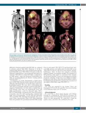
Figure 3. Images of a 63-year old female patient with histologically confirmed extramedullary (EM) manifestation of acute myeloid leukemia (AML) in the oral cav- ity (dotted arrows) who underwent intensive induction chemotherapy. (A) Maximum intensity projection (MIP) and (B) fused multiplanar reconstruction (MPR) of the pre-therapeutic 18Fluorodesoxy-glucose positron emission tomography/computed tomography (18FDG-PET/CT). Maximum standardized uptake value (SUVmax) was 9.1. 18FDG-PET detected a further right iliac EM AML (SUVmax 5.6; continuous arrows). (C) MIP of the post-therapeutic follow up 18FDG-PET/CT confirming the slightly regressive EM AML of the oral cavity (SUVmax 7.4) but also the progressive right iliacal EM AML (SUVmax 8.1). (D) MPR of this scan. New bicervical EM AML (dashed arrows) was also detected (E), see also (C) (SUVmax up to 9.5).
differences between patients with EM AML as compared to patients without EM AML. However, 3 of the 9 relapsed AML patients who were included in the trial har- bored EM AML as per 18FDG-PET/CT. In spite of this, the National Comprehensive Cancer Network and others rec- ommend 18FDG-PET/CT when EM AML is suspected in an AML patient, while the European LeukemiaNet pro- vides no recommendations for doctors treating patients with EM AML.2,6,21,22
In summary, our study demonstrates a higher preva- lence of EM AML than previously reported and assumed, while the clinical prevalence was in the range of previous- ly published reports. We were able to demonstrate that 18FDG-PET/CT is feasible and safe in patients with AML at diagnosis. Furthermore, we were able to confirm that, in most patients, more than one metabolically active EM AML manifestation could be detected with 18FDG- PET/CT. Interestingly, six patients who were PET-positive at baseline and who had histologically confirmed EM AML were in hematologic CR but still had detectable 18FDG-avid EM AML depicting heterogeneity after treat- ment (Online Supplementary Figure S1A). When analyzing those four patients who presented with EM AML at diag-
nosis (as per initial 18FDG-PET/CT) and frank bone mar- row AML, all four were still positive for EM AML as per 18FDG-PET/CT but in a CR as per bone marrow aspirate; furthermore, three of these four patients eventually relapsed, despite consolidation therapy. The dynamics of PET-positivity with regard to concurrent bone marrow involvement, therefore, seem to be heterogeneous and the prognostic relevance of this has to be studied in further prospective trials. Hence, this study allows further trials to be designed and calculated for a prospective evaluation of the impact of EM AML using 18FDG-PET/CT.
Funding
This study was supported by the German Cancer Aid Foundation (109575 to FS, KZ, and MS). Open Access funding by the Publication Funds of the TU Dresden.
Acknowledgments
We would like to thank Katrin Rosenow and Gabriele Kotzerke for co-ordinating the 18FDG-PET/CT procedures. We would like to thank Annett Engmann, Katrin Peschel, and Catrin Theuser for data management. We would like to thank the patients, nurses, and doctors who contributed to this study.
haematologica | 2020; 105(6)
1557
