
Page 287 - Haematologica - Vol. 105 n. 6 - June 2020
P. 287
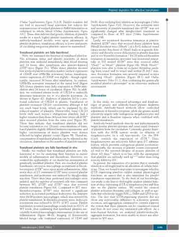
 Platelet depletion for effective transfusion
   (Online Supplementary Figure S2A-B). Platelet isolation did not lead to increased basal activation but reduces the responsiveness of washed platelets to PAR4 stimulation as compared to whole blood (Online Supplementary Figure S2C). These data indicate that genetic ablation of platelets results in a much higher platelet transfusion efficacy. In antibody-mediated models, even upon adjustment of platelet depletion, transfusion efficacy is lower and purity of circulating exogenous platelets cannot be maintained.
Transfused platelets are fully functional
Next, we examined the function of transfused platelets. The activation status and platelet reactivity of donor platelets was analyzed immediately after blood drawing and 24 hours after transfusion into platelet-depleted iDTRPlt mice (Figure 5C). While donor platelets showed low levels of platelet activation marker (surface expression of CD62P and GPIIb/IIIa activation) before transfusion, surface expression of CD62P was slightly - though signif- icantly- increased 24 hours after transfusion. In contrast, GPIIb/IIIa activation remained at the initial level (Figure 5D). Donor platelets were fully responsive to PAR4 stim- ulation after 24 hours of circulation (Figure 5D). In addi- tion, we evaluated plasma levels of CXCL4 to indirectly determine indications for in vivo platelet activation. As shown in Figure 4D, depletion of platelets led to a pro- found reduction of CXCL4 in plasma. Transfusion of platelets increased CXCL4 concentration although it did not reach basal levels, which can be explained by the reduced number of platelets. Noteworthy, basal CXCL4 levels with each mouse’s endogenous platelets, had a higher variation than those 24 hours later where all iDTRPlt mice received platelets from the same pool (Figure 5E). These data indicate that transfused platelets did not become activated in vivo. Of note, concentrations of trans- fused platelets slightly differed between experiments, and higher concentrations of donor platelets were always reflected by higher platelet counts (Figure 5F). Therefore, concentration of transfused platelets can be adjusted in the circulation, dependent on the number of platelets injected.
Transfused platelets are fully functional in vivo
Finally, we verified that transfused platelets are fully functional in vivo by analyzing their function in mouse models of inflammation and thrombosis. Moreover, we verified the applicability of our model for examination of genetically modified platelets. First, we checked if platelet transfusion can rescue macrophage recruitment in throm- bocytopenic iDTRPlt mice upon sterile inflammation. After seven days of DT treatment iDTRPlt mice received platelet transfusion, and peritonitis was induced by thioglycollate injection. Three days later, peritoneal macrophages were isolated, and the number of recruited cells was compared between WT, iDTRPlt and iDTRPlt mice that received platelet transfusion (Figure 6A). Compared to WT mice, thrombocytopenic iDTRPlt mice showed a significant reduction in recruited peritoneal leukocytes; the majority were F4/80 positive macrophages, which was restored by platelet transfusion. In thrombocytopenic mice, leukocyte recruitment was reduced to 30.9% of WT counts. Platelet transfusion reverted macrophage counts back to 92.1% of WT levels, thus confirming that transfused platelets suc- cessfully fostered macrophages extravasation to the site of inflammation (Figure 6B-D). Imaging of fluorescently labeled lavage cells confirmed expression of CD45 and
F4/80, thus verifying their identity as macrophages (Online Supplementary Figure S3A). Moreover, the activation state and reactivity of platelets transfused into iDTRPlt was not significantly changed after thioglycollate treatment as compared to those of WT mice (Online Supplementary Figure S3B).
Lastly, we monitored thrombus formation in platelet- depleted iDTRPlt mice that received donor platelets from Nbeal2 knockout mice (Nbeal2-/-) in a FeCl3-induced vessel injury model. Knockout of Nbeal2 leads to α-granule defi- ciency and thereby severe deficiencies in platelet function and in vivo thrombus formation.13 FeCl3 induced thrombus formation in mesenteric arterioles was monitored intravi- tally in DT treated iDTRPlt mice that received either Nbeal2+/+ or Nbeal2-/- platelets (Figure 6E). While Nbeal2+/+ platelets were able to form an occlusive thrombus reach- ing from vessel wall to vessel wall within 21.4±8.7 min- utes, thrombus formation was severely impaired in mice receiving Nbeal2-/- platelets (Figure 6F-G and Online Supplementary Video S1-2), thus confirming the genetically modified platelet’s phenotype14 in an otherwise unaltered environment.
Discussion
In this study, we compared advantages and disadvan- tages of genetic and antibody-based platelet depletion methods. Antibody-based methods allow controlled adjustment of thrombocytopenia, while the genetic model selectively targets only endogenous megakaryocytes and platelets and is therefore superior when combined with platelet transfusion.
Antibody-based methods directly and indiscriminately target murine platelet epitopes and cause rapid clearance of platelets from the circulation. Contrarily, genetic deple- tion with the iDTR system works via ablation of megakaryocytes, as a cell type-specific Cre, the PF4- iCre4, controls the expression of iDTR.15 Thus, megakaryocytes are selectively killed upon DT adminis- tration, which prevents endogenous platelet production. Additionally, the decrease of platelet counts correspond- ed well to the reported lifespan of murine platelets of about 4.8 days,10 which is in line with the assumption that platelets are naturally used up7,16,17 rather than being actively killed by DT.
In general, the expression of a protein that is normally absent in a cell might lead to an alteration of cellular func- tions. Thus, we found it important to investigate whether iDTR expressing platelets exhibit normal physiological functions, an aspect that is also important for platelet transfusion experiments. To the best of our knowledge, our study is the first to thoroughly analyze platelet func- tion of mice expressing iDTR in megakaryocytes and thus also on the platelet surface. We tested the classical platelet activators thrombin and collagen, as well as ago- nists that selectively trigger P2Y1/12, PAR4 and GPVI recep- tors. The fact that iDTR-expressing platelets did not show any noteworthy difference in activation, granule secretion, and aggregation compared to controls supports the notion that these platelets can be considered normal and thus do not cause any unwanted side-effects. As a non-classical function, we analyzed platelet-leukocyte- aggregate formation, but were unable to detect any alter- ations to WT mice.
haematologica | 2020; 105(6)
  1745
