
Page 255 - Haematologica May 2020
P. 255
leukins 5 and 7 in other cells,10-12 the proposed mechanism associating DNM2 loss-of-function mutations and ETP- ALL development.5 Further, previous studies using phar- macological approaches have suggested that dynamin GTPase activity contributes to receptor desensitization in human platelets, as in the case of the purinergic receptors P2Y1 and P2Y12.13
Human platelets express all three classical dynamins,14,15 including an inactive DNM3 spliced variant, for which a single nucleotide polymorphism has been associated with platelet size.16 In comparison, mouse platelets express pre- dominantly the ubiquitous DNM2,14,17 thus providing a valuable model to study DNM2-dependent RME in platelet and MK biology, independent of neuronal DNM1 and DNM3. We have previously shown that Dnm2fl/fl Pf4- Cre (Dnm2Plt–/–) mice specifically lacking DNM2 in the platelet lineage develop severe macrothrombocytopenia due to membrane fission arrest and accumulation of clathrin-coated vesicles obstructing the MK demarcation membrane system, the highly organized membrane reser- voir for future platelets.9 Here we investigated the role of DNM2 in platelet hemostatic function using both phar- macological and genetic approaches. Our data show that DNM2 regulates proximal signaling via the platelet colla- gen receptor GPVI and that DNM2-dependent RME is required for the accumulation of plasma fibrinogen into α-granules to facilitate normal platelet hemostatic func- tion.
Methods
Mice
Dnm2Plt–/– mice were described previously.9 Mice were treated according to the National Institutes of Health and Medical College of Wisconsin Institutional Animal Care and Use Committee guidelines.
Platelet count
Platelet count was measured on a Sysmex XT-2000i automatic hematology analyzer using blood collected by mouse retro- orbital plexus bleeding and immediately diluted in Cellpack (Sysmex) supplemented with EDTA and PGE1.18
Tail bleeding time
Bleeding time was determined by snipping 2 mm of distal mouse tail and immediately immersing the tail in 37°C isotonic saline.19 A complete cessation of bleeding was defined as the bleeding time.
Ex vivo perfusion assay
Platelet interaction with immobilized type I collagen was per-
formed using the VenaFlux Platform and Vena8Fluor+ biochips (Cellix).20 Additional information can be found in the Online Supplementary Methods.
Platelet preparation and flow cytometry
Blood was collected by mouse retro-orbital plexus bleeding and was anticoagulated in acid-citrate-dextrose.19 Platelets were isolated by sequential centrifugation, resuspended at 5x10⁸ platelets/mL, and incubated for 30 minutes (min) at 37°C with 100 μM of the non-competitive inhibitor of dynamin GTPase activity, dynasore (EMD Millipore),13,21-23 or vehicle (0.1% DMSO).
Platelets were activated or not with collagen-related peptide
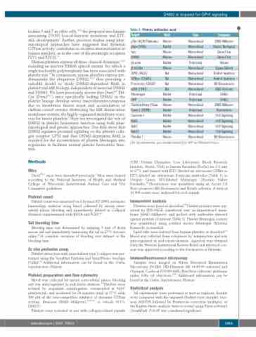
Table 1. Primary antibodies used.
DNM2 is required for GPVI signaling
Target
pTyr (4G10 Platinum)
Host
Mouse
Rabbit Mouse Mouse Rabbit Mouse Rat Rat Rat Rat Rabbit Rabbit Mouse Rabbit Rabbit Rabbit Rabbit Rabbit Mouse
Type
Monoclonal
Monoclonal Monoclonal Monoclonal Polyclonal Monoclonal Monoclonal Monoclonal Monoclonal Monoclonal Polyclonal Polyclonal Monoclonal Polyclonal Monoclonal Monoclonal Monoclonal Monoclonal Monoclonal
Company
EMD Millipore
Boster Biological Santa Cruz Santa Cruz Abcam Sigma-Aldrich Emfret Analytics Emfret Analytics BD Biosciences R&D Systems DAKO DAKO EMD Millipore Proteintech Cell Signaling Cell Signaling Cell Signaling Cell Signaling BD Biosciences
pLyn (Y396) Lyn
DNM2
β-actin
β-tubulin
GPVI (JAQ1) GPIbα (CD42b) P-selectin (CD62P) αIIb (CD41) Fibrinogen
vWF
Clathrin Heavy Chain Cavin 2 (SDPR) Caveolin 1
Rab5
Rab7
Rab11
Flotillin 1
haematologica | 2020; 105(5)
pTyr: phosphotyrosine; pLyn: phosphorylated Lyn; vWF: von Willebrand factor.
(CRP; Protein Chemistry Core Laboratory, Blood Research Institute, Versiti, USA) or human thrombin (Roche) for 2-3 min at 37°C and stained with FITC-labeled rat anti-mouse GPIbα or FITC-labeled rat anti-mouse P-selectin antibodies (Table 1) or Oregon Green 488-labeled fibrinogen (Thermo Fisher Scientific).19 Fluorescence was quantified using an Accuri C6 flow cytometer (BD Biosciences) and FlowJo software. A total of 10,000 events were analyzed for each sample.
Immunoblot analysis
Platelets were lysed as described.19 Platelet proteins were sep- arated by SDS-PAGE, transferred onto an Immobilon-P mem- brane (EMD Millipore), and probed with antibodies directed against proteins of interest (Table 1). Platelet fibrinogen content was quantitated using purified mouse fibrinogen (Enzyme Research) as standard.
Lipid rafts were isolated from human platelets as described.24 Blood was collected from volunteers by venipuncture and was anticoagulated in acid-citrate-dextrose. Approval was obtained from the Western Institutional Review Board and informed con- sent was approved according to the Declaration of Helsinki.
Immunofluorescence microscopy
Samples were imaged on Nikon Structured Illumination Microscopy (N-SIM, NIS-Elements AR v4.40.00 software) and Olympus Confocal FV1000-MPE (FluoView software) platforms under 100x oil objectives.9,25 Additional information can be found in the Online Supplementary Methods.
Statistical analysis
All experiments were performed at least in triplicate. Results were compared with the unpaired Student t-test (simple), two- way ANOVA followed by Bonferroni correction (multiple), or the Kaplan-Meier analysis (time-to-event) using Prism software (GraphPad). P<0.05 was considered significant.
1415
