
Page 248 - Haematologica April 2020
P. 248
D.E. van der Wal et al.
A
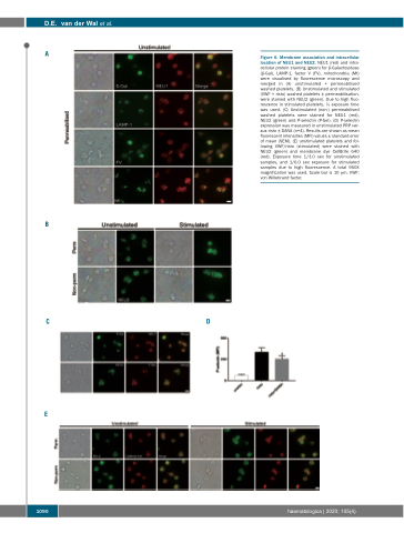
Figure 6. Membrane association and intracellular location of NEU1 and NEU2. NEU1 (red) and intra- cellular protein staining (green) for β-Galactosidase (β-Gal), LAMP-1, factor V (FV), mitochondria (Mt) were visualised by fluorescence microscopy and merged in (A) unstimulated + permeabilised washed platelets. (B) Unstimulated and stimulated (VWF + risto) washed platelets ± permeabilisation, were stained with NEU2 (green). Due to high fluo- rescence in stimulated platelets, 1⁄2 exposure time was used. (C) Unstimulated (non-) permeabilised washed platelets were stained for NEU1 (red), NEU2 (green) and P-selectin (P-Sel). (D) P-selectin expression was measured in unstimulated PRP ver- sus risto ± DANA (n=4). Results are shown as mean fluorescent intensities (MFI) values ± standard error of mean (SEM). (E) unstimulated platelets and fol- lowing VWF/risto (stimulated) were stained with NEU2 (green) and membrane dye CellBrite 640 (red). Exposure time 1/3.0 sec for unstimulated samples, and 1/6.0 sec exposure for stimulated samples due to high fluorescence. A total 960X magnification was used. Scale bar is 10 μm. VWF: von Willebrand factor.
B
CD
E
1090
haematologica | 2020; 105(4)
