
Page 167 - Haematologica April 2020
P. 167
CD99 overexpression in AML
CD99 monoclonal antibody treatment exhibits antileukemiaactivityinacutemyeloidleukemiacells
Acute myeloid leukemia patient blasts (n=7) incubated with monoclonal CD99 antibody (mAbCD99: 20 μg/mL)
showed a significant decrease in cell viability (48 h: 0.5±0.02-fold, P<0.0001) (Figure 7A). Treatment with mAbCD99 caused a decrease in colony formation in 3 of 6 patient samples: AML-2 (0.5-fold, P=0.02), AML-6 (0.65-
AB
CDE
FGH
IJ
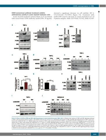
Figure 8. Effect of CD99 on ERK and SRC signaling pathways in acute myeloid leukemia (AML) cells. (A) Western blot analysis of SRC and ERK signaling pathways in THP-1 cells transduced with EV, CD99-L, and CD99-S lenti-virus for 72 hours (h). (B) Western blot analysis of SRC and ERK signaling pathways in THP-1 cells trans- fected with siCD99 for 48 h. (C) MDM2 western blot analysis in THP-1 cells transduced with EV, CD99-L, and CD99-S lentivirus. (D) MDM2 analysis performed by western blot in THP-1 cells treated with mAbCD99 for 1, 2, and 9 h. (E) ERK signaling pathway analyzed in THP-1 cells treated with mAbCD99 for 1, 3, and 9 h. (F) Cyclin D1 mRNA expression measured by taqman assay in THP-1 cells transduced with EV, CD99-L and <CD99-S lentivirus. (G) CCND1 mRNA expression measured by taqman assay in THP-1 cells transfected with siCD99. (H) Western blot analysis of SRC and ERK signaling pathways in THP-1 stable cells transduced with EV, CD99-L and CD99-S lentivirus. (I) SRC signaling pathway analyzed in THP-1 cells treated with mAbCD99 for 1 and 9 h. (J) ERK and SRC signaling pathway analyzed in MOLM-13 cells treated with mAbCD99 for 1, 2, 6, and 9 h. (****P<0.0001; *P<0.05).
haematologica | 2020; 105(4)
1009
