
Page 14 - Haematologica April 2020
P. 14
Editorials
HIF-1α: a potential treatment target in chronic lymphocytic leukemia
Martina Seiffert
Molecular Genetics, German Cancer Research Center (DKFZ), Heidelberg, Germany E-mail: MARTINA SEIFFERT - m.seiffert@dkfz.de
doi:10.3324/haematol.2019.246330
In 2019, William Kaelin Jr., Peter J. Ratcliffe and Gregg L. Semenza were jointly awarded the Nobel Prize in Physiology or Medicine for their work in elucidating how the transcription factor Hypoxia-inducible factor 1 (HIF-1) senses oxygen availability and adapts cellular response accordingly.1 HIF-1 is a heterodimeric protein complex that consists of two proteins: a constitutively expressed HIF-1β subunit and an inducible HIF-1α sub- unit. During hypoxia, HIF-1α hydroxylation is reduced, preventing its proteasomal degradation, and the stabi- lized HIF-1 complex comprised of HIF1α and HIF-1β is transported to the nucleus where it regulates the expres- sion of several hundred genes to counter the lack of oxy- gen.
In solid cancer, hypoxia is widely involved in tumor biology. Overexpression of HIF-1α is associated with aggressive cancer cell behavior and is correlated with poor overall patient survival. Tumor cells react to low oxygen levels by inducing HIF-1α expression, which results in an activation of many crucial cancer hallmarks, such as angio- genesis, glucose metabolism, cell proliferation/viability, invasion and metastasis.2 Even though hypoxia was ini- tially identified as a driver of HIF-1α expression, it has become clear in recent years that its overexpression in cancer can be also driven by genetic alterations, such as gain-of-function mutations in oncogenes or loss-of-func- tion mutations in tumor-suppressor genes.
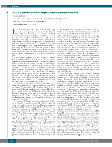
In this issue of Haematologica, Griggio et al. report that disrupting mutations in the tumor-suppressor gene TP53 are associated with higher HIF-1α levels and activity in patients with chronic lymphocytic leukemia (CLL).3 They further show that elevated HIF-1α levels are due to an increased transcription as well as a decreased protein degradation of HIF-1α. Evidence for both regulatory mechanisms have already been described in CLL and are summarized in Figure 1.
A constitutively higher expression and activity of HIF- 1α in CLL cells compared to normal B cells was suggested to be due to reduced levels of the von Hippel-Lindau gene product (pVHL), which is responsible for HIF-1α degrada- tion. Downregulation of pVHL was attributed to the inhibitory activity of miR-155, a highly abundant microRNA in CLL which is suggested to contribute to increased HIF-1α stability in CLL cells.4 As pVHL expres- sion levels were lower in CLL patients with disrupted TP53 compared to wild-type TP53 cases, Griggio et al. sug- gest that the observed accumulation of HIF-1α protein in TP53-disrupted CLL might be partially explained by an increased stability of HIF-1α.
It has recently been suggested that the mutant forms of FBXW7 contribute to HIF-1α stability in CLL cells.5 FBXW7 is known to target proto-oncoproteins including HIF-1α for proteasomal degradation. In silico modeling predicted that mutations in FBXW7 that recurrently occur
in CLL change the binding of protein substrates including HIF-1α and are therefore involved in the increased stabili- ty of HIF-1α in patients with these mutations.
HIF-1α has been suggested to be transcriptionally regu- lated in CLL cells via signals delivered by the microenvi- ronment. In oxygenated blood, circulating CLL cells were shown to be primed for hypoxia and to undergo a rapid induction of HIF-1α activity when entering lymphoid tis- sues.6 In these tissues, CLL cells are in constant interaction with accessory cells which deliver essential signals for sur- vival and proliferation of CLL cells and contribute to resistance to drug-induced apoptosis.7 The contact of CLL cells with stromal cells induces HIF-1α expression via induction of phosphatidylinositol 3-kinase (PI3K) and ERK mitogen-activated protein kinase (MAPK) signaling. In addition, hypoxia in lymphoid tissues was shown to mod- ify central metabolic pathways in CLL cells, including increased adenosine generation and signaling via the A2A adenosine receptor.8 Inhibiting the A2A receptor has been shown to counteract the effects of hypoxia in CLL and accessory cells, and to increase their susceptibility to phar- macological agents.
In line with this, Griggio et al. observed increased expression of HIF-1α in CLL cells upon co-culture with stromal cells to a level that is comparable to hypoxic con- ditions.3 They further showed that HIF-1α transcript lev- els of primary CLL cells positively correlate with CLL cell survival in culture. This is likely related to the observation that HIF-1α critically regulates the interaction of CLL cells with their microenvironment. It has previously been shown that HIF-1α induces an upregulation of chemokine receptors, such as CXCR4, and cell adhesion molecules that control the interaction of leukemic cells with bone marrow (BM) and lymphoid microenvironments.9 In line with this, inactivation of HIF-1α was shown to impair chemotaxis and cell adhesion to stroma, reduce BM and spleen colonization in xenograft and allograft CLL mouse models, and prolong survival of these mice. In support of a modulation of CLL cell motility, HIF-1α transcript levels were shown to correlate with the expression of CXCR4 and other target genes in CLL patient samples.
Further evidence for stroma-induced activation of HIF- 1α in CLL comes from a comparative transcriptome analy- sis of primary CLL cells co-cultured or not with protective BM stromal cells which revealed that oxidative phospho- rylation, mitochondrial function and hypoxic signaling are the most significantly deregulated functions in non-pro- tected CLL cells.10 These relevant transcriptomic changes were compared to the Connectivity Map database,11 which contains transcriptomic responses of cell lines to treatment with small molecules. This comparison identi- fied drugs that act by repressing HIF-1α and disturbing intracellular redox homeostasis as potential compounds blocking stroma-mediated support of CLL cells.
856
haematologica | 2020; 105(4)
