
Page 138 - Haematologica April 2020
P. 138
H. Kumar et al.
A
B CDE
H
FG
IJKL
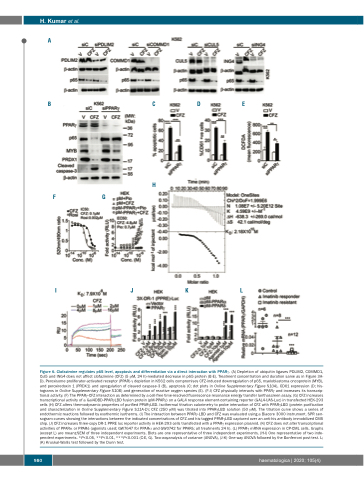
Figure 6. Clofazimine regulates p65 level, apoptosis and differentiation via a direct interaction with PPARγ. (A) Depletion of ubiquitin ligases PDLIM2, COMMD1, Cul5 and ING4 does not affect clofazimine (CFZ) (5 μM, 24 h)-mediated decrease in p65 protein (B-E). Treatment concentration and duration same as in Figure 3A- D). Peroxisome proliferator-activated receptor (PPAR)-γ depletion in K562 cells compromises CFZ-induced downregulation of p65, myeloblastoma oncoprotein (MYB), and peroxiredoxin 1 (PRDX1) and upregulation of cleaved caspase-3 (B), apoptosis (C; dot plots in Online Supplementary Figure S10A), CD61 expression (D; his- tograms in Online Supplementary Figure S10B) and generation of reactive oxygen species (E). (F-J) CFZ physically interacts with PPARγ and increases its transcrip- tional activity. (F) The PPARγ-CFZ interaction as determined by a cell-free time-resolved fluorescence resonance energy transfer lanthascreen assay. (G) CFZ increases transcriptional activity of a Gal4DBD-PPARγLBD fusion protein (pM-PPARγ) on a GAL4 response element-containing reporter (GAL4-UAS-Luc) in transfected HEK-293 cells (H) CFZ alters thermodynamic properties of purified PPARγLBD. Isothermal titration calorimetry to probe interaction of CFZ with PPARγLBD (protein purification and characterization in Online Supplementary Figure S11A-D); CFZ (250 μM) was titrated into PPARγLBD solution (50 μM). The titration curve shows a series of endothermic reactions followed by exothermic isotherms. (I) The interaction between PPARγ-LBD and CFZ was evaluated using a Biacore 3000 instrument. SPR sen- sogram curves showing the interactions between the indicated concentrations of CFZ and his-tagged PPARγLBD captured over an anti-his antibody immobilized CM5 chip. (J) CFZ increases three-copy DR-1 PPRE luc reporter activity in HEK-293 cells transfected with a PPARγ expression plasmid. (K) CFZ does not alter transcriptional activities of PPARα or PPARδ (agonists used; GW7647 for PPARα and GW0742 for PPARδ; all treatments 24 h). (L) PPARγ mRNA expression in CP-CML cells. Graphs (except L) are mean±SEM of three independent experiments. Blots are one representative of three independent experiments. (H-I) One representative of two inde- pendent experiments. *P<0.05, **P<0.01, ***P<0.001 (C-E, G). Two-wayanalysis of variance (ANOVA), (J-K) One-way ANOVA followed by the Bonferroni post-test. L; (K) Kruskal-Wallis test followed by the Dunn test.
980
haematologica | 2020; 105(4)
