
Page 121 - Haematologica April 2020
P. 121
TM+ monocytes in myelodysplastic syndromes
the Dunn multiple comparisons test was used. A Spearman corre- lation was computed for PB and BM comparisons. P values <0.05 were considered statistically significant. Graphpad Prism 6 soft- ware (San Diego, CA USA) was used for graphic display and sta- tistical calculations. A multivariate Cox regression analysis for overall and leukemia-free survival was performed using IBM SPSS Statistics software version 22 (New York, NY, USA).
Results
Classical monocytes in patients with myelodysplastic syndromes express thrombomodulin
Monocyte subsets were identified by flow cytometric analysis based on the expression of CD14, CD16 and M- DC8 according to recently published recommendations.38 Classical, intermediate and non-classical monocytes were characterized by using the above mentioned markers (Figure 1A). Subsequently, the expression of TM and HLA- DR, a major histocompatibility complex (MHC) molecule class II, was assessed on all monocyte subsets derived from normal bone marrow (NBM) and MDS BM samples. Monocyte subsets from MDS patients showed high levels of TM expression on their cell surface, whereas NBM- derived monocytes showed very low levels of TM. HLA- DR expression, too, was higher on all monocyte subsets in MDS BM than it was in NBM (Figure 1A). The levels of expression on different monocyte subsets were quantified in a larger cohort of patients (NMB samples, n=10 and MDS BM samples, n=24). Total percentages of monocyte subsets in MDS BM compared to NBM were not signifi- cantly different. However, the percentage of monocytes that expressed TM was significantly higher for MDS- derived classical monocytes compared to the same mono- cyte subset in NBM (37.3% vs. 9.9%; P<0.0001) (Figure 1B). The percentages of TM expression on intermediate and non-classical monocytes were equally distributed between MDS BM and NBM (Figure 1B). The median flu- orescent intensity of TM and HLA-DR was evaluated on the three distinct monocyte subsets using the same set of samples. Classical monocytes from MDS BM showed higher levels of TM expression compared to classical monocytes fron NMB (3.7-fold higher; P<0.0001). HLA- DR expression levels were higher for all MDS-derived monocyte subsets (classical monocytes: 2.0-fold, P=0.0015; intermediate monocytes: 1.8-fold, P=0.0154; non-classical monocytes: 1.5-fold, P<0.0001) (Figure 1B). TM expression remained unchanged upon overnight stim- ulation with TLR ligands in a preliminary set of samples (Online Supplementary Figure S2).
Classical monocytes were then analyzed in a larger set of samples, since this subset forms the most prevalent subset in NBM as well as in MDS BM, and revealed the most prominent difference in TM expression. The PB compartment was also included in the analysis. The cohort was extended with 130 MDS BM-derived samples and 15 NBM samples. The PB and BM samples in the extended control and patient cohorts (normal PB, n=31; MDS PB, n=29; NBM, n=25 and MDS BM, n=154) were screened for the presence of TM on classical monocytes (Figure 1C). MDS-derived classical monocytes in both the PB and BM compartment showed higher expression of TM compared to the expression on classical monocytes from normal PB and NBM samples (PB: 33.6% vs. 17.8%, P=0.015; BM: 37.0% vs. 8.6%, P<0.0001). Furthermore, a
strong positive correlation was found for the percentage of classical monocytes expressing TM in the two compart- ments in 25 paired MDS samples (r=0.83, P<0.0001) (Figure 1C). The same flow cytometric panel was used to study TM expression on other cell types present in the PB and BM,. We were able to identify granulocytes, eosinophils and B cells. TM was exclusively expressed on MDS-derived monocytes (Online Supplementary Figure S3) and none of the other cell types in PB and BM showed positivity for TM, including the non-B cell lymphocytic compartment consisting of T and NK cells (data not shown).
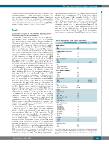
Table 1. Characteristics of the patients and controls.
Characteristics
Total number
Value
[min-max]
[45-85]
HD 56
MDS 183
Peripheral blood samples
Number 60 HD 31 MDS 29
Age – mean, years
HD -
MDS
69
Sex
HD – male/female - MDS – male/female 20/9
Bone marrow samples
Number 179 HD 25 MDS 154
Age – mean,y HD
62 [20-86]
110/44
MDS
69 [36-94] HD – male/female 18/7
Sex
MDS – male/female
IPSS-R
Very low risk
Low 20 Low risk 34 Intermediate risk 22 High risk 8 Very high risk 8
WHO classification
MDS-SLD 5 MDS-MLD 48 MDS-RS-SLD 8 MDS-RS-MLD 27 MDS-EB1 22 MDS-EB2 20 Del5q 12
% Blasts
<5% 97 ≥5% 44
Ring sideroblasts
No 106 Yes 40
HD: healthy donor; MDS: myelodysplastic syndrome; IPSS-R: Revised International Prognostic Scoring System; WHO, World Health Organization; SLD, single lineage dys- plasia; MLD, multilineage dysplasia; RS, ring sideroblasts; EB, excess blasts; Del5q: dele- tion 5q.
haematologica | 2020; 105(4)
963
