
Page 93 - Haematologica March 2020
P. 93
ELANE knockout restores granulopoiesis in congenital neutropenia
ical translational start site and/or internal start sites in exon 2 are disrupted and that expression of internally-ini- tiated ELANE is pathogenic. We found a marked reduction of ELANE mRNA, most probably due to the induction of nonsense-mediated mRNA decay (NMD) of ELANE mRNA after exposure of cells to ELANE-specific CRISPR/Cas9 sgRNA RNP. We also did not detect any additional NE protein bands on WB analysis of edited cells using antibody recognizing C-terminus of NE. Based on these observations, we concluded that our sgRNA is inducing loss of NE protein without activation of the path- ogenic ELANE forms from the internal ATG.
We did not detect off-target activity in edited cells, but recent results from Alan Bradley have suggested that the introduction of CRISPR/Cas9 editing can cause multiple genomic changes far beyond the actual target.28 Therefore,
A
for clinical applications, evaluation of the off-target activi- ty of CRISPR/Cas9 on whole genome level using next- generation sequencing should be performed. In addition, it would be important to evaluate that the editing of ELANE occurred in the repopulating hematopoietic stem cell pop- ulation and that these cells maintained their ability to engraft immunodeficient mice in vivo. Since most probably HSC are not expressing NE, we will not expect any dam- aging effects of the ELANE KO on the functions and integrity of HSC.
We demonstrated here, that CRISPR/Cas9 mediated ELANE KO in HSPC and iPSC of CN patients induces granulocytic differentiation and in vitro generated ELANE KO neutrophils have no defects in the phagocytic activity, ROS production, and chemotaxis. NE is a proteolytic enzyme of the neutrophil serine protease (NSP) family,
B
C
D
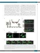
Figure 8. Human ELANE KO PMN are capable to migrate and to phagocyte S. aureus BioParticles in zebrafish embryos in vivo. (A) The scheme of in vivo phagocytosis assay in zebrafish embryos xenotransplanted with human fluorescently labeled polymorphonuclear leukocytes (PMN). (B) A representative confocal image highlight- ing the presence of transplanted human ELANE KO PMN in the caudal hematopoietic site of a zebrafish embryo at 53 hpf. White arrows indicate S. aureus BioParticles phagocytosed by human ELANE KO PMN. CV: caudal vein; DA: dorsal aorta. Scale bar: 20 mm. (C) Three-dimensional rendering of a z-stacks of 12 mm illustrating human ELANE KO PMN, one of them has engulfed S. aureus BioParticles (white arrow). Scale bar: 10 μm. (D) Still photographs from a time-lapse recording illustrating the phagocytic activity of transplanted human ELANE KO PMN in the zebrafish embryo. Arrowheads indicate the formation of neutrophil protrusions. Numbers indicate time in minutes. Scale bar: 10 mm.
haematologica | 2020; 105(3)
607
