
Page 302 - Haematologica March 2020
P. 302
M. Berger et al.
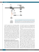
Figure 7. Proposed signaling pathway downstream of CD36 through which oxidized low-density lipopro- teins (oxLDL) activate phosphodiesterase 3A (PDE3A) to suppress cyclic adenosine monophosphate (cAMP) signaling. OxLDL binds to CD36 to activate a tyrosine kinase signaling pathway that leads to a sus- tained activation of PDE3A. Under these conditions cAMP is maintained at low concentrations through hydrolysis to 5’AMP (solid red line), and the activation of PKA is diminished (dashed red line). Through dis- inhibition, this pathway acts to reduce the threshold for platelet activation and promote thrombosis.
of FA6-152 (1 mg/mL), but not control IgG (1 mg/mL), caused a significant recovery in phosphoVASP-ser157 (Figure 3A and B). OxPCCD36 (25 mM), but not the con- trol lipid, PAPC (25 mM), diminished phospoVASP-ser157 in response to PGI2 (Figure 3C). Thrombin-induced aggre- gation of wild-type (WT) and CD36-/- platelets was indis- tinguishable, and and PGI2 (20 nM) caused complete inhi- bition of aggregation with only shape change remaining (Figure 3D and E). However, in CD36-deficient platelets, oxPCCD36 (25 mM) did not influence the inhibitory effects of PGI2 (Figure 3E). To confirm the mechanism underpinning these observations, we examined cAMP sig- naling. PGI2 (20 nM) caused a significant increase in cAMP in both WT and CD36-/- platelets (4382±175 and 4128±366 fmol cAMP/1x108 platelets). Preincubation with oxLDL, but not nLDL, reduced cAMP accumulation in WT mice (3347±294 fmol cAMP/1x108 platelets; P<0.05) but not CD36-/- (4196±224fmol cAMP/1x108 platelets) (Figure 3F). Furthermore, PGI2-induced phosphoVASP-ser157 (Figure 3G) and ser239 (Online Supplementary Figure S5) were reduced by oxLDL in WT but not CD36-/- platelets.
Atherogenic lipid stress induces platelet phosphodiesterase 3A activity through a mechanism that requires CD36, Src kinases and protein kinase C
Our data suggested that ligation of CD36 could activate PDE3A. We tested this directly measuring enzymatic activity of PDE3A immunoprecipitated from platelets treated with either oxLDL or oxPCCD36. PDE activity in the immunoprecipitated samples was blocked by milri- none confirming enzyme activity was due to PDE3A (Online Supplementary Figure S6). OxLDL (10-100 mg/mL) caused a concentration-dependent increase in PDE3A activity (Figure 4A, left), which plateaued at 50 mg/mL (24±6.8%; P<0.05 compared to basal) and was maintained
at 27.7%±8.5 above basal for up to 60 min (longest time tested) (Figure 4A, right). This was strikingly different from the physiological agonist thrombin which induced a rapid induction of PDE3A activity that peaked at 1 min before returning to basal after 5 min (Online Supplementary Figure S7).
To link the sustained activation of PDE3A to decreased cAMP levels, we assessed the cyclic nucleotide concentra- tions over time. Platelets were incubated with oxLDL for up to 60 min, followed by a 1 min treatment with PGI2 (50 nM) before measuring cAMP concentrations. The pre- incubation of platelets with oxLDL, but not nLDL (data not shown), for up to 60 min significantly blunted PGI2-induced increases in cAMP with concentrations remaining at basal levels for the time course (Figure 4B). To confirm the role of CD36 in the activation of PDE3A, experiments were repeated with oxPCCD36. In human platelets, oxPCCD36 (25 mM), but not PAPC (25 mM), increased PDE3A activity (26.9±7.3%; P<0.05 compared to basal and PAPC) (Figure 4C). Critically, oxPCCD36 stimulated activity in WT platelets to 25.4±2.4% (P<0.05 compared to basal or PAPC) but not in CD36-/- platelets (Figure 4D).
We previously described a CD36-specific signaling pathway that includes the sequential activation of Src- family kinases (SFK), Syk, PLCγ2 and protein kinase C (PKC)17 and investigated whether these kinases were involved in the activation of PDE3A. The non-selective SFK inhibitor, dasatinib (10 mM), ablated oxPCCD36- induced PDE3A activation (Figure 4E), while the Syk inhibitor, R406 (1 mM), caused significantly reduced PDE3A activity (Figure 4F). Given that CD36 signaling leads to PKC activation in a SFK manner, and that PDE3A is a target of PKC in platelets,31 we tested the PKC inhibitors RO318220 (10 mM) and BIM1 (10 mM), and the intracellular Ca2+ chelator BAPTA-AM (20 mM) (Figure
816
haematologica | 2020; 105(3)
