
Page 297 - Haematologica March 2020
P. 297
CD36 activates platelet PDE3A
inhibition was unaffected by nLDL (5.8±1.2%). Similar data were obtained when platelets were stimulated with collagen (Online Supplementary Figure S1). Next, the effects of oxLDL in whole blood were examined. Stimulation of whole blood with PAR-1 peptide (10 mM) increased P-selectin expression, which was reduced by pre-treat- ment with PGI2 (20 nM). Consistent with the aggregation experiments, oxLDL reduced inhibitory effects of PGI2 (Figure 1B). Finally, the effects of oxLDL under physiolog- ical conditions of arterial flow were examined. Perfusion of whole blood over collagen-coated biochips led to platelet deposition and thrombus formation (Figure 1C), with surface coverage inhibited by PGI2 (20 nM) from 14.9±2.9 to 3.8±1.7% (P<0.05). OxLDL, but not nLDL, prevented PGI2 -mediated inhibition of platelet deposition (8.1±1.9%; P<0.05 compared to PGI2 alone). OxLDL alone caused a small but non-significant increase in thrombosis and was incorporated into the thrombi as evidenced by staining for oxidized lipid epitopes (Figure 1D). These data demonstrate that under a variety of different conditions oxLDL reduces platelet sensitivity to PGI2.
Oxidized low density lipoproteins modulate cyclic adenosine monophosphate signaling through increased phosphodiesterase 3A activity
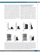
PGI2 inhibits platelets through the stimulation of cAMP- PKA signaling cascade.20 Given the reduced platelet sensi- tivity to PGI2, the direct effect of oxLDL on platelet cAMP metabolism was tested. Incubation with PGI2 caused a sig- nificant increase in platelet cAMP concentrations (1814±166 fmol cAMP/1x108 platelets; P<0.05 vs. basal). When platelets were treated with nLDL (50 mg/mL), the ability of the prostanoid to elevate cAMP was unaffected (1885±203 fmol cAMP/1x108 platelets), while oxLDL (50 mg/mL) prevented PGI2-induced accumulation of cAMP (481±23 fmol cAMP/1x108 platelets; P<0.05 com- pared to PGI2 alone) and also reduced basal cAMP concen- trations (not significant) (Figure 2Ai). To determine if oxLDL blocked cAMP synthesis or accelerated cAMP breakdown by phosphodiesterase 3A (PDE3A) and PDE225,25 the PDE2A inhibitor EHNA (20 mM) and the PDE3A inhibitor milrinone (10 mM) were used. Consistent with previous studies, both inhibitors potentiated cAMP
AB
CD
Figure 2. Oxidized low-density lipoproteins modulate cyclic adenosine monophosphate (cAMP) signaling in response to prostacyclin (PGI2). (A) Washed human platelets (2x10⁸/mL) incubated with apyrase, indomethacin and EGTA were treated alone or with control native LDL (nLDL) or oxLDL (50 mg/mL) for 2 minutes (min) followed by a 1 min PGI2 (50 nM) incubation. Platelets were lysed and intracellular cAMP concentrations were measured by enzyme immunoassay. (Left) Intracellular cAMP levels presented as mean±standard error of mean (SEM) (n=3, *P<0.05, Mann-Whitney U Test). (Center) Platelets treated as described under (A) in the pres- ence EHNA (20 mM). Intracellular cAMP levels are presented as mean±SEM (n=3, *P<0.05, Mann-Whitney U Test) (Right) Platelets treated as described under (A) in the presence of Milrinone (10 mM). Intracellular cAMP levels are presented as mean±SEM (n=3, *P<0.05, Mann-Whitney U Test). (B) Washed human platelets (2x10⁸/mL) were incubated alone or with oxLDL (50 mg/mL) for 2 min followed by a 5-min incubation with Forskolin (10 mM), then lysed and measured as described under (A). Intracellular cAMP concentrations presented as mean±SEM (n=3, *P<0.05, Mann-Whitney U Test). (C) Washed human platelets (5x10⁸/mL) were treated as described under (A), lysed in Laemmli buffer, separated by SDS-PAGE and immunoblotted with anti-phosphoPKA substrate, anti-phosphoVASPser157 and anti-β tubulin. (Left) Representative blot of three independent experiments. (Right) Densitometry of the representative highlighted band (red) (*P<0.05, Mann-Whitney U Test). (D) Washed human platelets were treated as described under (A) except that platelets were treated with 8-CPT-cAMP (50 mM) for 5 min and then processed as in (C). (Left) Representative blot of three independent experiments and (Righti) densitometry of the representative highlighted band (red).
haematologica | 2020; 105(3)
811
