
Page 296 - Haematologica March 2020
P. 296
M. Berger et al.
A
B
C
DE
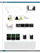
Figure 1. Oxidized low-density lipoproteins (oxLDL) induce prostacyclin (PGI2) hyposensitivity in platelets. (A) Washed human platelets (2.5 x 10⁸/mL) were treated alone or with control native LDL (nLDL) or oxLDL (50 mg/mL) for 2 minutes (min) followed by a 1 min incubation with PGI2 (20 nM). Thrombin (0.05U/mL)-stimulated aggregation was then measured under constant stirring (1000 rpm) at 37°C for 4 min. In some cases, platelets were incubated with either thrombin, nLDL or oxLDL alone for 4 min. (Left) Representative aggregation traces. (Right) Percentage aggregation is presented by mean±standard error of mean (SEM) (n= 5). **P<0.01 compared to platelets treated with thrombin and PGI2, Mann-Whitney U Test. (B) Whole blood was treated as in (A) and activated with PAR-1 peptide (10 μM) for 5 min followed by fixation. CD62P expression was assessed by flow cytometry. Representative data of three independent experiments. (Left) Data are presented as heatmap of mean fluorescence intensity (MFI). (Right) Representative histograms. (C) Human whole blood was incubated with PGI2 (20 nM) for 1 min alone or with control native LDL (nLDL) or oxLDL (50 mg/mL) for 10 min. Blood was perfused over collagen-coated biochips for 2 min at arterial shear 1000s-1 and images of adherent platelets were taken by fluorescence microscopy. (Left) The surface coverage (%) is presented as mean±SEM (n=3) (P<0.05, Mann-Whitney U Test). (Right) Representative fluorescence microscopy images are shown. (D) Whole blood was incubated with nLDL or oxLDL (50 mg/mL) for 10 min and then perfused over col- lagen-coated surfaces for 2 min at arterial shear 1000s-1. Thrombi were post-stained with anti-oxPC antibody and images were taken by fluorescence microscopy. (Left) Representative of images of 3 independent experiments. Stained with DIOC6 (top panel) or anti-oxPC (bottom panel) (Center) The surface coverage is present- ed as mean±SEM (n=4, *P<0.05, Mann-Whitney U Test). (Right) Fluorescence (red Channel) is presented as mean±SEM (n=4, *P<0.05, Mann-Whitney U Test).
810
haematologica | 2020; 105(3)
