
Page 288 - Haematologica March 2020
P. 288
M. Ando et al.
A
BC
D
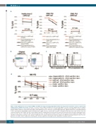
Figure 3. Induced pluripotent stem cell-derived LMP1– and LMP2-rejT show cytotoxicity against Epstein-Barr virus-infected-infected tumors equal to or greater than original cytotixic T lymphocytes in vitro. (A) In vitro 51Cr release assay of LMP1- and LMP2-rejT (effectors) and original CTL clones (effectors) against auto LCL (targets) and HLA-mismatched LCL (control targets). (B) Flow cytometric analysis of PD-1 expression of original LMP2-CTLs and iPSC-derived LMP2-rejT. (C) Flow cytometric analysis of PD-L1 and PD-L2 expression on the ENKL cell line NK-YS. (D) In vitro 51Cr release assay of original LMP2-CTL, LMP2-rejT (effectors), and HLA-mismatched T cells (control effector) against ENKL (targets). Anti-PD-L1 Ab(+), ENKL cells were cultured with 10 mg/mL of anti-PD-L1 antibody for three days until the assay was conducted. Anti-PD-L1 Ab(-), ENKL cells were cultured without anti-PD-L1 antibody. Data are presented as mean ± SD and represent at least three independent exper- iments. E:T ratio: effector : target ratio; iPSC: induced pluripotent stem cell; LMP: latent membrane protein; rejT: rejuvenated cytotoxic T lymphocytes; ENKL: extran- odal NK/T-cell lymphoma nasal type; CTL: cytotoxic T lymphocytes; Pt: patient; HLA: human leukocyte antigen; LCL: EBV-infected lymphoblastoid cells; PD-1: pro- grammed cell death 1; PD-L1: programmed death-ligand 1; PD-L2: programmed death-ligand 2; Ab: antibody; SD: standard deviation.
802
haematologica | 2020; 105(3)
