
Page 286 - Haematologica March 2020
P. 286
M. Ando et al.
HLA-A*02– tumors. These results indicated that LMP1- specific and LMP2-specific rejT generated from multiple donors, including an ENKL patient, had strong antigen- specific cytotoxicity against ENKL.
LMP1-specific and LMP2-specific rejT tended to show stronger cytotoxicity against EBV+ tumor cells than the original CTL clone in vitro
We next compared rejT cytotoxicity against EBV+ tumor cells with that of original CTL by 51Cr release assay. A*2402-restricted LMP2-rejT generated from a healthy donor more efficiently killed autologous EBV-infected LCL (70.4% and 65.4%; E:T ratios of 20:1 and 10:1) compared to the original CTL clone (51.7% and 49.4%; E:T ratios of 20:1 and 10:1). A*0201-restricted LMP1-rejT generated from an ENKL patient showed somewhat stronger cyto- toxicity against autologous EBV-infected LCL (90.3%, 90.0%, 77.8%, and 58.8%; E:T ratios of 40:1, 20:1, 10:1 and 5:1) than the original CTL clone (77.8%, 61.9%, and 43.8%; E:T ratios of 20:1, 10:1 and 5:1). The cytotoxicity of A*0201-restricted LMP2-rejT against autologous EBV- infected LCL (44.0% and 34.5%; E:T ratios of 40:1 and 20:1) was almost the same as that of the original CTL clone (40.4% and 33.7%; E:T ratios of 40:1 and 20:1). We further compared PD-1 expression of peripheral blood- derived original EBV-CTL and of rejT from the same LMP2-CTL clone, measured by flow cytometry: values were 15.3% for EBV-CTL and undetectable for rejT (Figure 3B). PD-L1 expression of ENKL cells, measured by flow cytometry, was 57.4% (Figure 3C). Although both LMP2-specific rejT and original LMP2-CTL (PYLFWLAAI) powerfully killed HLA class I-matched NK-YS cells, rejT cytotoxicity tended to be stronger (60.8%, 52.6%, 52.6%, and 40.1%; E:T ratios of 40:1, 20:1, 10:1 and 5:1 specific 51Cr release, respectively) than that of the original CTL (59.7%, 50%, 40.9%, and 39.8%; E:T ratios of 40:1, 20:1, 10:1 and 5:1 specific 51Cr release, respectively) (Figure 3D). To elucidate whether PD-L1 blockade can enhance the potential to kill ENKL of the original CTL that express PD- 1+ and of rejT that do not express PD-1–, ENKL cells were cultured with 10 mg/mL of anti-PD-L1 antibody for three days immediately preceding the assay. Anti-PD-L1 anti- body did not clearly enhance killing by either PD-1+ origi- nal CTL or PD-1– rejT (Figure 3D). Our results demonstrat- ed definite cytotoxic activity against EBV-infected tumor
cells in vitro for both EBV-specific original CTL and rejT, with cytotoxicity of rejT against ENKL cells stronger than that of original CTL and without killing enhancement in original CTL or rejT cultured with anti-PD-L1 antibody.
Robust anti-ENKL effect and marked survival improvement of LMP2-rejT in vivo
ENKL cells express not only PD-L1 but also PD-L2 (Figure 3C). Anti-PD-1 antibody that can block both PD- L1 and PD-L2 was thus potentially more effective than anti-PD-L1 antibody as therapy for ENKL. Using PD-1 blockade to observe whether EBV-specific rejT exert ENKL-suppressive effects in vivo and anti-PD-1 antibody has an additive antitumor effect, ENKL cells labeled with retrovirus-derived firefly luciferase were intraperitoneally engrafted into NOG mice (1x10⁵ cells/mouse). Light emis- sion was monitored as an indicator of tumor growth. Four days after tumor inoculation, these mice were divided into two control groups and four treatment groups. No treat- ment was given in one control group (n=6) and only anti- PD-1 antibody was given in the other (200 mg per dose, three doses) (n=6). The treatment groups consisted of mice treated with original LMP2-CTL (5x10⁶ per dose, three doses) (n=6); mice treated with original LMP2-CTL
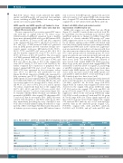
Table 2. PD-L1+, PD-L1+/–, and PD-L1– extranodal NK/T-cell lymphoma nasal type: patient characteristics.
6
and anti-PD-1 antibody (5x10 of original CTL and 200 μg
of anti-PD-1 antibody per dose, three doses) (n=6); mice treated with LMP2-specific rejT (5x10⁶ per dose, three doses) (n=6); and mice treated with LMP2-specific rejT and anti-PD-1 antibody (5x10⁶ of rejT and 50 mg of anti- PD-1 antibody per dose, three doses) (n=5).
By day 21, bioluminescence had progressively increased in the control-group mice (no treatment, 176.8-fold, range 7.33-326.0; anti-PD-1 Ab, 53.3-fold, range 15.5-101.0) (Figure 4A-B). In contrast, tumor suppressive effects were observed in the original CTL group (6.025-fold, range 1.27-10.4) and in the original CTL with anti-PD-1 Ab group (17.5-fold, range 0.71-51.1), with and even stronger suppressive effects in the rejT group (0.505-fold, range 0.31-0.816) as well as in the rejT with anti-PD-1 Ab group (0.71-fold, range 0.50-1.10). Tumor signals regressed fur- ther in the four groups treated with original CTL, original CTL with anti-PD-1 antibody, rejT, or rejT with anti-PD-1 antibody than in the untreated group (original CTL, P=0.0001; original CTL with anti-PD-1 antibody, P=0.0003; rejT, P<0.0001, and rejT with anti-PD-1 anti-
PD-L1 + N=3 mean/N
48.00
PD-L1 +/- N=8 mean/N SD/%
52.43 20.35
2/8 25.0% 5/8 62.5% 2/5 40.0% 5/8 62.5% 6/8 75.0% 2/3 66.7%
57.14 44.69
PD-L1 - N=17
P value 0.945
1.000 0.566 0.707 1.000 0.590 0.038
Age
Sex
Advanced
EBV DNA-positive SMILE
CR
LMP1
SD/%
23.30
mean/N
52.14
5/17
11/17
8/8
9/17
9/17
5/6
44.77
SD/%
20.12
29.4%
64.7%
100.0%
52.9%
52.9%
83.3%
41.48
1/3 33.3% 3/3 100.0% 3/3 100.0% 1/3 33.3% 0/3 0.0% 0/3 0.0%
Observation period (month) 5.67 6.43
Programmed death ligand-1 (PD-L1); ENKL: extranodal NK/T-cell lymphoma,nasal type;EBV: Epstein-Barr virus;SMILE: dexamethasone,methotrexate,ifosfamide,L-asparaginase and etoposide; CR: complete remission; PD-L1: programmed death-ligand 1; LMP: latent membrane protein; SD: standard deviation.
800
haematologica | 2020; 105(3)
