
Page 175 - Haematologica March 2020
P. 175
KS99 alone or in combination in AML
ABC
DE
FG
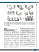
Figure 1. KS99 inhibits cell proliferation and clonogenicity and induces apoptosis in human acute myeloid leukemia (AML) cell lines. (A) Cell viability of AML cell lines after the treatment with KS99. (B) IC50 values for AML cell lines were plotted with 95% confidence intervals. (C) Induction of apoptosis with KS99 was determined as the percentage of Annexin V-positive cells. (D) The sensitivity of human AML cell lines to KS99 or Cytarabine (Ara-C) alone. (E) OCI-AML3 and MV4-11 cells were treated with increasing doses of KS99, Ara-C or combination. (E, left) Combination Index (CI) values of KS99 and Ara-C co-treatment were calculated by CalcuSyn. Synergy CI<0.9. (E, right) Apoptosis was determined as the percentage of Annexin V-positive cells. (F and G) KS99 reduced the colony-forming ability of AML cell lines. The representative colony microscopy images (4X) are shown as indicated. Data are the mean±standard error of the mean. *P<0.05; **P<0.01; ***P<0.001; ****P<0.0001; one-way ANOVA.
Results
KS99 induces apoptosis and reduces cell survival of human acute myeloid leukemia cell lines
To assess the effect of KS99 on AML cells in vitro, a panel of human leukemic cell lines [MOLM-13, MV4-11, OCI- AML2, OCI-AML3, HL-60, vincristine resistant HL-60 (HL-60/VCR), U937, and KG-1], and mouse leukemia C1498 cells were selected. Cell viability was measured after treatment with KS99 (10 nM-10 mM) for 48 hours (h). For most of the cell lines, KS99 treatment led to a decrease in the viability in the nanomolar (nM) range (Figure 1A). The half inhibitory concentration (IC50) values for all the human cell lines were between 100 nM and 600 nM. Specifically, MV4-11, MOLM-13, and OCI-AML2 mani- fested higher sensitivity with lower IC50 values of 166 nM, 228 nM, and 218 nM, respectively. The other cell lines showed relatively higher IC50 values (300-600 nM) (Figure 1B). Similarly to human AML cells, mouse C1498 cells were sensitive to KS99 with an IC50 of 217 nM.
Furthermore, flow cytometry demonstrated apoptosis on selected cell lines. KS99 induced apoptosis within the nanomolar range (100-500 nM) in a dose-dependent man- ner (Figure 1C). MOLM-13 cells were chosen for time- dependent survival, and the outcome showed an early decrease in cell proliferation at 6-8 h with a prominent decline in viability post 24-48 h of treatment (Online Supplementary Figure S1A). Simultaneously, apoptosis was evident as early as 4 h of treatment with maximum effect at 48 h (Online Supplementary Figure S1B). These time
points were considered when further functional assays were conducted. Chemotherapeutics are clinically often used in combination to achieve complete remission (CR) in AML patients. Hence, we decided to compare and com- bine KS99 to Ara-C to evaluate an increase in efficacy of Ara-C. The sensitivity of AML cell lines to KS99 and Ara- C is shown in Figure 1D. For the synergy studies, OCI- AML3 and MV4-11 cells were treated with Ara-C (0.062- 4 mM) and KS99 (0.03125-2 mM) for 72 h, and the combi- nation index (CI) was calculated. KS99 significantly increased cytotoxic responses, in combination with Ara- C, and showed synergy (CI<0.9) in both cell lines (Figure 1E, left panel). Furthermore, subtoxic KS99 concentrations (lower than IC50) reduced IC50 of Ara-C by a median of 2- 3-fold (Online Supplementary Figure S1C). Similarly, KS99 augmented the pro-apoptotic effect of Ara-C in OCI- AML3 and MV4-11 cells (Figure 1E, right panel). Next, the inhibitory effect of KS99 on colony forming ability of human AML cell lines was determined. The treatment of KS99 led to a decrease in the number and size of colonies across the selected range and cell lines (Figure 1F and G). These results show that KS99 is active in AML and can be combined with Ara-C to enhance anti-leukemic activity.
KS99 exerts a cytotoxic effect in primary human acute myeloid leukemia and favors cases with poor prognosis The pro-apoptotic activity of KS99 was tested on newly
diagnosed and untreated primary human AML patients (n=21). Cells were treated with increasing concentrations of KS99 (0.1-6 mM) for the 48 h. The IC50 values were cal-
haematologica | 2020; 105(3)
689
