
Page 162 - Haematologica March 2020
P. 162
M. Xu et al.
(Ensembl GRCh37 release 92). We evaluated the function of the hBCR/ABL1 oncoprotein in zebrafish by over- expressing hBCR/ABL1 mRNA encoding the p210BCR/ABL1 oncoprotein. We then detected the numbers of myeloid cells during embryonic hematopoietic development by
AB
CD
EF
GH
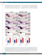
lcp1, lyz, mpx whole-mount in situ hybridization (WISH), and Sudan Black B (SB) cytochemical staining (Figure 1). lcp1, also named l-plastin, is a pan-myeloid marker that identifies all myeloid subsets, including macrophages and neutrophils. Numbers of lcp1+ cells were significantly
IJKL
Figure 1. Expression levels of myeloid markers (lcp1, lyz, mpx and SB) increased after transient expression of humanized BCR/ABL1 (hBCR/ABL1) in zebrafish lar- vae. Whole mount in situ hybridization (WISH) of lcp1 (A and B), lyz (C and D), and mpx (E and F) expressions in wild-type (WT) zebrafish larvae after transient over- expression of hBCR/ABL1 mRNA (right) were higher than controls (left) at 3 dpf. (G and H) The number of SB+ cells in WT zebrafish larvae after transient overexpres- sion of hBCR/ABL1 mRNA (right) was higher than controls (left) at 3 dpf. n/n: number of zebrafish larvae showing representative phenotype/total number of zebrafish larvae examined. Original magnification ×32 (A-H). Red rectangles in each panel indicate the signals in PBI region, and the regions were enlarged at the lower right (original magnification ×200). (I-L) Statistical analysis. lcp1+ signals (I), lyz+ signals (J), mpx+ signals (K), and SB+ signals (L) in PBI region after injection were calculated and compared at 3 dpf (Student t-tests, mean±Standard Error of Mean, **P<0.01; ****P<0.0001).
676
haematologica | 2020; 105(3)
