
Page 115 - Haematologica March 2020
P. 115
Novel PDE9i therapy for sickle cell disease
in SCD leads to multi-organ pathology. To assess the impact of IMR-687 on vessel occlusion, HbSS-Townes mice were exposed to 1 h of hypoxia (7% O2/ 93% N2) and the percentage of static venules (no blood flow) was quantified after return to normoxic conditions using a DSFC and intravital microscopy. After vehicle-treated mice were returned to normoxia, microvascular stasis was 33% and 16% after 1 h (Figure 4A) and 4 h (Figure 4B), respectively. Treatment with IMR 687 for ten days decreased stasis to 12% (P<0.01 vs. vehicle) and 7% (P<0.01) at 30 mg/kg/day and 20% (P<0.05) and 14% (ns) at 10 mg/kg/day after 1 h and 4 h in normoxia. Treatment of SS mice with HU at 100 mg/kg/day for ten days decreased microvascular stasis to 13% (P<0.05) and 8% (P<0.05) after 1 h and 4 h, respectively. When mice were given the combination of IMR 687 (30 mg/kg/day) and HU (100 mg/kg/day), stasis was 7% (P<0.01) and 4% (P<0.01) at 1 h and 4 h, respectively, suggesting a potential synergistic effect of the two agents.
Fetal hemoglobin induction in sickle cell disease patient erythroblasts
Erythroblasts were generated in vitro using two-phase liquid culture from CD34+ progenitors from nine SCD blood or bone marrow (SCD patients undergoing hip replacement for osteonecrosis) donors. These cells were treated with IMR-687 to determine if the drug could increase HbF expression in patient-derived erythrocytes.
F-cells were determined by their expression of HbF in the LiveDead-GPA+Band3+ population (Figure 5) by FACS. The mean for the DMSO control group (n=9) was 13.3% HbF positive. IMR-687 increased the percentage of F-cells to 21.9% (P<0.01, n=9). HU increased the percentage of F- cells to 22.2% (P<0.01, n=7, due to cytotoxicity induced by HU in 2 cultures). HU had a greater impact on the
intensity of HbF staining in blood-derived CD34+ cells, increasing the MFI of the cells to 9744±2805 compared to 6073±1217 in control cells (P=0.041, n=7, due to cytotox- icity in 2 cultures), while IMR-687 significantly increased the MFI to 7813±1374 (P<0.01, n=9). This difference may be due in part to the greater cytotoxic stress of culturing the cells in 30 mM HU, evidenced by the loss of 2 of the 9 HU cultures.
Phosphodiesterase 9 inhibitor IMR-687 demonstrated low central nervous system accumulation and did not alter behavior
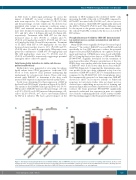
Many PDE9i were originally developed for neurological diseases.49-55 In contrast, IMR-687 is a novel PDE9i selected specifically for low CNS exposure to reduce the potential impact of neuronal PDE9 inhibition on cognitive develop- ment and function. C57Bl/6J mice were dosed with IMR- 687 at 10 mg/kg/day for five days or a CNS-active PDE9i, PF-04447943, originally developed for the treatment of neurological disorders. Plasma concentrations of the two PDE9i were very similar, while the brain exposure levels of IMR-687 were 5-fold lower than those seen with PF- 04447943 (Figure 6A). Comparing the brain/plasma expo- sure profiles of the two drugs confirmed a very low con- centration of IMR-687 in the CNS (7% brain/plasma ratio) compared to the PF-04447943, (41% brain/plasma ratio). Not unexpectedly, given its low brain exposure, IMR-687 showed no effect on locomotor activity or behavioral responses in toxicology studies (data not shown) nor in a classical fear conditioning mouse model of learning and memory (Figure 6B) (see Online Supplementary Methods). In contrast, the brain penetrant PF-04447943 significantly increased conditioned fear responses in mice at a similar dose. Besides confirming the lack of CNS activity of IMR- 687, this finding suggests that brain-penetrant PDE9i treat-
AB
Figure 6. A brain-penetrant phosphodiesterase-9 inhibitor (PDE9i), but not IMR-687, increases fear responses in a model of learning and memory. (A) Fear condi- tioning responses are increased and persistent in mice treated with a brain-penetrant PDE9i compared to vehicle-treated or IMR-687-treated mice. (B) Drug exposure of the brain-penetrant PDE9i is 5-fold greater than that of IMR-687. Errors are presented as Standard Error. ns: not significant.
haematologica | 2020; 105(3)
629
